

Quick take: Most skin bumps like these are harmless, but you should watch for changes.
If you have any of these signs, contact your doctor:
• The bump grows quickly or gets larger than expected.
• It starts to bleed, hurt, or change shape.
• You see a sudden color change or irregular border.
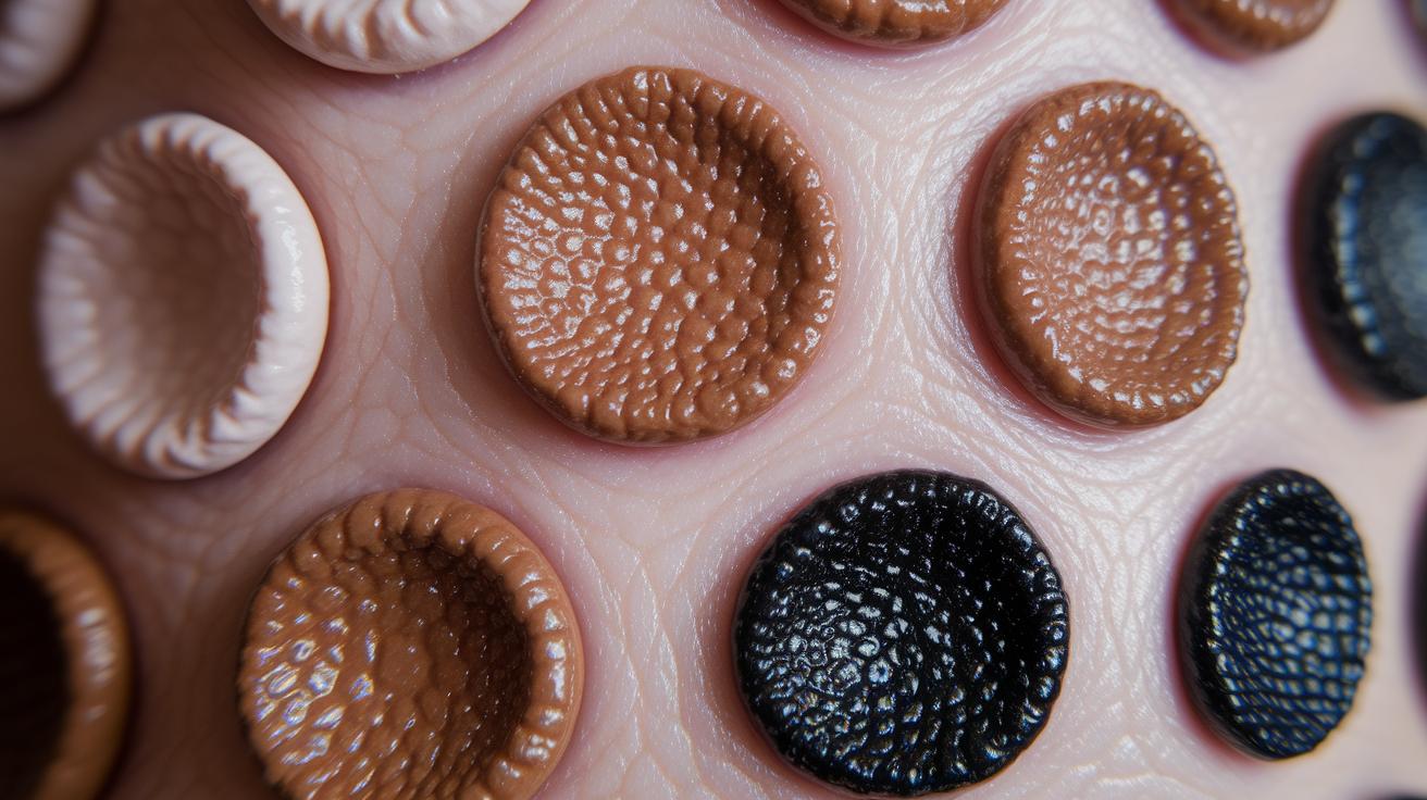
This gallery shows clear, high-quality images of seborrheic keratoses (common, usually non-cancerous skin spots). You may see bumps that differ in color, size, and texture. Look at these pictures to learn what you might expect and to help you track any shifts over time. Knowing the differences can guide you on when it might be a good idea to consult your doctor. Our guide is here to give you clear, trustworthy information so you feel confident about caring for your skin.
Seborrheic keratosis pictures Shine with Visual Clarity
This gallery shows clear, high-quality pictures of common skin bumps called seborrheic keratoses. These harmless growths come in different sizes, colors, and textures. Knowing what they normally look like can help you track any changes.
- Image 1: A small bump (papule) that is skin-colored on the chest. The close-up shows a mildly rough edge that looks as if it is simply attached to the skin.
- Image 2: A flat, raised spot (plaque) that is tan and appears on the back. The detailed view shows a well-defined texture.
- Image 3: A small bump (papule) with a light brown color on the shoulder. The close-up brings out a fine, grainy surface.
- Image 4: A flat lesion (plaque) that is dark brown on the back. The close-up shows a thick and uneven texture with a clearly attached look.
- Image 5: A skin-colored bump (papule) on the chest. The close-up captures tiny irregular features and a slightly crumbly edge.
- Image 6: A black flat lesion (plaque) on the shoulder. The detailed view reveals a rough and scaled surface.
- Image 7: A tan small bump (papule) on the back. The close-up highlights a smooth border with a subtle waxy finish.
- Image 8: A dark brown flat spot (plaque) on the chest. The detailed view shows an irregular, firm look that clearly seems stuck on.
Even though these growths look different from one another, they are all benign and do not turn into cancer. Keeping an eye on these features can help you know when it might be time to talk with a doctor about any changes.
Seborrheic Keratosis Picture Variations in Color and Shape
Below is a gallery that shows different types of seborrheic keratoses. These are common, non-cancerous skin spots that can vary in color, size, and texture. This clear breakdown helps you compare them easily.
| Appearance | Color | Size | Surface |
|---|---|---|---|
| Skin-colored papule | Skin-colored | <1 cm | Waxy, smooth |
| Brown plaque | Dark brown | 1–2 cm | Rough, stuck-on |
| Black crusty lesion | Black | >2 cm | Crumbly, scaly |
| Pink flat patch | Pink | <1 cm | Flat, smooth |
This combined view makes it easy to spot the differences in each type of lesion, so you can better understand their unique features and know what to look for.
Seborrheic Keratosis Picture Locations on the Body
On your chest and back, seborrheic keratoses usually appear as rough, stuck-on spots. In image 1, you see a skin-colored lesion with a firm, rough edge on the chest. In image 4, a dark brown patch with an uneven surface appears on the back. These examples show how SK often looks on larger sections of skin.
On your face and scalp, these growths tend to look lighter and more subtle. Image 2 shows a lightly pigmented lesion on the face, while image 3 shows a similar patch on the scalp where the skin is thinner. This uniform look is different from the rugged texture seen on the torso.
In rare cases, SK can develop on very sensitive areas like the eyelids. Image 8 shows a small, delicate lesion with soft borders on a sensitive facial spot. In these areas, be sure to keep an eye on any changes since the skin is thinner and more delicate.
Comparative Seborrheic Keratosis Pictures and Other Lesions

Comparing these skin spots helps you keep track of changes and know when to see a doctor.
• SK versus actinic keratosis: SK shows a smooth, sleep-on look with one color and no bleeding. Actinic keratosis (sun-damaged spot) looks rough and scaly and may turn into cancer.
• SK versus melanoma: Melanoma usually has uneven borders and mixed colors and may bleed or develop an ulcer. In contrast, SK keeps a steady color with a smooth, clear edge and does not bleed.
• Texture differences: SK spots can feel smooth and waxy or a little rough. Actinic keratosis feels scaly from sun damage, while melanoma may feel irregular.
• Color and border clues: SK has even color and sharp borders. Other spots, like melanoma, have mixed colors and jagged edges that could signal something more serious.
If you see a sudden change like the spot getting larger, causing pain, or itching, especially with a color change, please see a doctor right away.
Seborrheic Keratosis Close-up Picture Textures
Close-up images let you see the tiny details that help doctors confirm that seborrheic keratoses (harmless skin spots) are benign. They show you the lesion’s texture and clear borders, making it easier to tell these spots apart from others that might need more attention.
Macro photos work like a magnifying glass. They reveal little features such as milia-like cysts (small white bumps) and comedo-like openings (dark spots from keratin buildup). You might see surfaces that range from flaky keratin plugs to a smooth, waxy layer that looks stuck on. These details are key for a safe and accurate check.
When closely examined, the well-known "stuck-on" edge is clear. This reassures clinicians that the growth is non-cancerous. In short, these images help experts study every nuance, supporting careful skin checks and guiding safe care decisions.
Clinical and Dermoscopic Seborrheic Keratosis Pictures
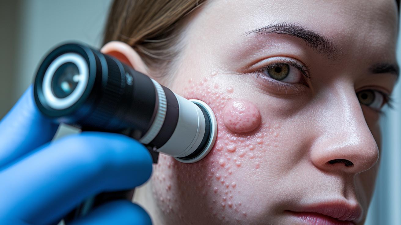
Dermoscopy is a tool that helps doctors closely look at seborrheic keratoses. It shows small details that might not be visible to the naked eye. For example, it can reveal tiny white spots called milia-like cysts, fine cracks, and little dark dots (comedo-like openings) from built-up skin. One image might show a smooth, even surface highlighted by these tiny cysts under high magnification.
Reflectance confocal microscopy uses lasers to capture cell patterns directly on your skin. This technique lets doctors see how cells are arranged in the area. Computer-aided skin image analysis then measures features like edge smoothness and evenness of color. These details add objectivity to what might be hard to see with regular photos.
Using both dermoscopy and these advanced imaging methods, clinicians can be sure that seborrheic keratoses are harmless. They combine the close-up pictures with precise measurements to decide if treatments like cryosurgery or shave excision are needed when the lesion causes symptoms.
Final Words
In the action, you’ve seen a detailed guide on seborrheic keratosis images, covering a photo gallery, color and texture variations, anatomical locations, and differential comparisons. The post breaks down key points like texture details, clinical imaging, and red-flag signs that call for a review by a clinician. You now have clear guidance on what to watch for and track when comparing seborrheic keratosis pictures. Keep this info handy, stay observant, and remember that early checks help keep skin health on track.
FAQ
What does seborrheic keratosis removal cream do?
A seborrheic keratosis removal cream targets the lesion’s appearance by softening and gradually reducing the growth’s texture; however, it may not fully remove the lesion and it’s best to consult a clinician for proper treatment advice.
What do seborrheic keratosis NHS pictures and types of keratosis pictures show?
NHS and clinical images of seborrheic keratosis display stuck-on, rough lesions ranging from small papules to larger plaques with colors from skin-toned to dark brown or black, illustrating common benign features seen in people over 50.
How do pictures of actinic keratosis and seborrheic keratosis differ?
Images of actinic keratosis typically reveal rough, scaly patches caused by sun exposure while seborrheic keratosis pictures show a uniformly stuck-on appearance with smoother, well-defined borders and consistent color.
How do seborrheic keratosis pictures on the face appear?
Facial seborrheic keratosis images show lesions with a stuck-on appearance that may vary in color; these growths usually have clear, defined edges and occur more frequently in older adults.
What features are seen in pictures of large seborrheic keratosis and inflamed examples?
Large seborrheic keratosis pictures display plaque-type lesions over 1 cm with a rough or crumbly surface, while inflamed examples reveal redness and irritation suggesting mild inflammation around the growth.
How do seborrheic keratosis pictures on the scalp differ?
Scalp images of seborrheic keratosis typically show well-circumscribed lesions amid hair-bearing areas, often maintaining the classic stuck-on look, though their visibility might be affected by hair density.
What do seborrheic keratoses look like overall?
Overall, seborrheic keratoses appear as benign, stuck-on lesions with a rough or waxy texture, varying in size from small papules to larger plaques, and displaying colors such as skin-toned, tan, brown, or black.
How can one get rid of seborrheic keratosis naturally?
Natural remedies for seborrheic keratosis include options like apple cider vinegar or tea tree oil, though scientific support is limited; it’s crucial to consult a healthcare provider before starting any home treatment.
What triggers seborrheic keratosis formation?
Seborrheic keratosis is mainly triggered by aging and genetic predisposition, with some evidence that sun exposure and skin irritation might contribute; the lesions are benign and usually increase in number with age.
What differentiates actinic keratoses from seborrheic keratoses?
Actinic keratoses result from sun damage and appear as rough, scaly patches with a potential risk of progression, whereas seborrheic keratoses present a uniform, stuck-on look with no bleeding or rapid changes.