
Quick take: A skin spot might be harmless, but if it changes, it could need a check.
If you notice any of these emergency signs, call emergency services now:
• The spot grows a lot in a short time.
• It changes color or shape fast.
• It starts bleeding, hurting, or itching badly.
For spots that aren’t as urgent, here’s what to do:
• Look at the spot carefully. Has it stayed the same, or is it getting bigger or darker?
• Write down when you first saw it and any changes you notice.
• If it seems different from before or you feel worried, call your doctor or visit an urgent care center.
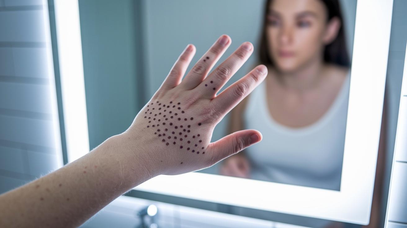
Most spots stay the same for many years. For example, a regular mole doesn’t change much. But a spot that starts shifting in size, shape, or color might be a sign of a skin issue.
Keep an eye on your skin. If you see red flags, don’t wait, get checked by a clinician right away.
Key Contrasts in Benign vs Malignant Skin Lesions
Quick take: Benign skin spots usually stay the same, but malignant ones change over time.
Triage Box: If you notice any of these signs, contact your doctor today:
- A new spot larger than 5 mm
- Uneven or jagged edges
- Mixed colors such as brown, black, red, or blue
- Changes like raised texture, bleeding, or itchiness
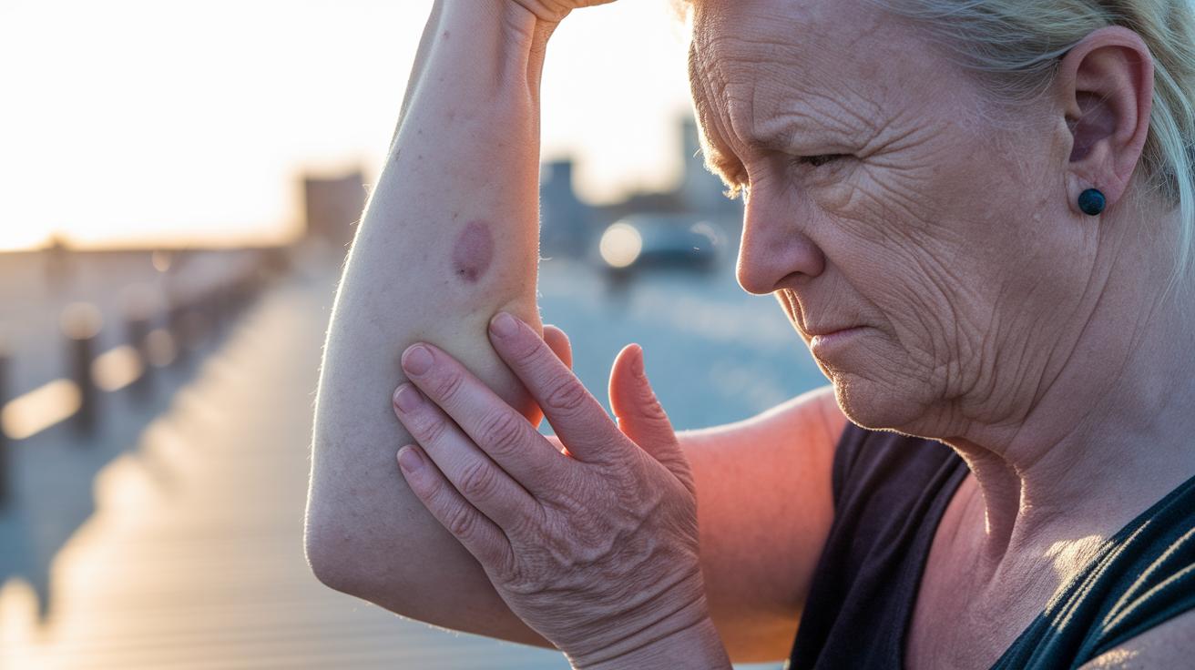
Benign lesions are non-cancerous spots that generally remain steady. They are often round or oval with smooth, clear borders and a uniform color. Many of these spots, like common moles, can fade or flatten as you age.
By contrast, malignant lesions tend to evolve over weeks or years. A spot that grows or changes, especially one over 5 mm, should be evaluated by a healthcare professional. These spots might have irregular shapes, fuzzy or jagged edges, and a mix of colors. They may also become raised, or even start bleeding or itching.
Keeping a simple log of any changes in your skin can help catch problems early. Regular self-exams are a smart way to monitor your skin and get help fast if a spot starts showing red-flag features.
ABCDE Criteria: Visual Features Differentiating Benign and Malignant Lesions
Quick take: Use these five simple checks to decide if a skin spot needs a doctor’s review.
If you notice any of these signs, get medical advice right away:
- The spot is changing quickly.
- The spot looks uneven.
- The edges are jagged or unclear.
- There are several colors in one spot.
- The spot is larger than 6 mm (about the size of a pencil eraser).
Most common moles are safe. They are even in shape, have smooth edges, show only one color, and remain stable over time. But if your spot shows any of the warning signs above, a doctor should take a look.
Here’s how to check your skin spot:
• A means asymmetry: One side does not mirror the other.
• B means border: Smooth edges are a good sign. Jagged edges can be a concern.
• C means color: One uniform color is usually safe, while mixed colors may signal a problem.
• D means diameter: Spots smaller than 6 mm tend to be benign. Spots over 6 mm or that are growing need a closer look.
• E means evolving: Changes in size, shape, or color should prompt a check-up.
| Criteria | Benign Feature | Concerning Feature |
|---|---|---|
| Asymmetry | Even, mirrored shape | Uneven, mismatched shape |
| Border | Smooth, clear edge | Jagged or blurred edge |
| Color | Uniform color | Multiple or mixed colors |
| Diameter | Under 6 mm | Over 6 mm or increasing |
| Evolving | Remains the same | Changes in size, shape, or color |
Keep these guidelines in mind during your skin checks. If any concerning signs appear, act now and consult a health professional.
Diagnostic Techniques for Benign vs Malignant Skin Lesions
Quick take: Skin exams catch small changes early.
If you notice any of these warning signs, call your doctor immediately:
- A spot that grows or changes very fast
- Different colors or blurry borders
- Bleeding or itching from a mole
Your doctor starts with a full-body skin check. They use special tools to look closely at your skin and compare each spot to your usual pattern. Even tiny differences in size, shape, or color can mean a mole is changing.
One important tool is dermatoscopy. This device gives a close-up view of your skin so the doctor can see tiny pigment patterns and blood vessels. They check if a mole looks even or if some areas are starting to look different. This helps tell apart harmless spots from ones that might need more attention.
Sometimes, more tests are needed. Your doctor may take a small piece of skin (a biopsy) to check it under a microscope. This test is the best way to confirm if a spot is cancer. In some cases, advanced imaging like confocal microscopy is used to get even more details. These extra tests help decide if a spot needs quick treatment.
Each step is part of a careful plan to decide the best treatment and keep you safe.
Risk Factors and Indicators for Malignant Skin Lesions
Sun damage is a major player in skin cancer risk. Too much ultraviolet light (sun or tanning beds) can harm your skin cells. If you have fair skin and have suffered blistering sunburns, your risk goes up. Changes in your skin, like a new spot or one that changes shape or color, should be checked quickly since they may be signs of basal cell or squamous cell cancer.
Family history also counts. If close family members have had skin cancer or if you carry certain genes, you are more at risk for melanoma (a serious skin cancer). That means any new or changing spot on your skin deserves attention. Keep a close eye on your skin and share any changes with your doctor right away.
When to Seek Evaluation for Benign and Malignant Skin Lesions
Checking your skin every month can help you catch changes early. By knowing what your skin normally looks like, you can spot any new changes more easily.
If you see any of the following red flags, act quickly:
• Uneven shape (one side does not mirror the other)
• Jagged or fuzzy edges
• Different colors within the same spot
• Growth beyond 5 mm (about the size of a pencil eraser)
• Changes such as itching, bleeding, or crusting
For instance, a mole with fuzzy edges might signal a concerning change. When you report these signs, share details about your sun exposure and family history. Your doctor may then suggest tests like dermatoscopy (a close-up exam of your skin) or a nearly painless biopsy.
Noticing these signs early helps you connect your self-checks with timely professional care.
Final Words
In the action, we've walked through key contrasts in skin lesion evaluation. We covered visual signs using the ABCDE rule, the role of professional screenings and biopsies, and environmental as well as genetic risk factors. The guide shows clear steps for self-monitoring and knowing when to seek urgent help. By focusing on differences between benign and malignant skin lesions, you can better track changes and discuss findings with your clinician. Stay alert and keep a positive view on managing your skin health.
FAQ
What does a types of skin lesions chart show?
A types of skin lesions chart shows different kinds of skin lesions, categorizing them by features such as size, shape, color, and border. It helps you quickly compare primary lesions with those that evolve secondarily.
What are primary and secondary skin lesions?
Primary lesions are the initial abnormal skin changes, while secondary lesions develop from the primary ones due to scratching, infection, or healing processes. This distinction aids in monitoring lesion evolution.
How do skin lesions pictures assist in understanding skin changes?
Skin lesions pictures provide visual references that make it easier to identify features like shape, color, and border irregularities in both benign and malignant lesions. They also help you track changes over time.
What can benign skin lesions pictures reveal?
Benign skin lesions pictures reveal uniform, smooth moles with consistent color and well-defined borders. They offer reassurance and serve as a baseline for noticing any future changes in the lesions.
What information do skin growths pictures provide?
Skin growths pictures display various types of lesions, highlighting differences in texture, color, and size. They can help you determine whether a growth appears non-cancerous or if further evaluation may be needed.
How is skin lesion treatment determined?
Skin lesion treatment is based on the lesion’s type and changes over time. Benign lesions are usually monitored, while suspicious or evolving lesions may require removal and biopsy. Always consult a dermatologist for personalized advice.