

Quick Take: A crater-like skin bump might be harmless or it could signal something that needs a closer look.
If you have any of these signs, call emergency services now:
• The bump grows quickly
• It starts bleeding or changes color
• It becomes painful or ulcerated
This type of bump often looks like a tiny volcano with a hardened center plug. It may be simple, but sometimes it requires treatment or further imaging tests (special scans to see inside your skin). One type, called keratoacanthoma (a fast-growing skin lesion), can change rapidly.
Keep an eye on your skin and note any changes in size, shape, or color so you can share accurate details with your doctor. I know it can be worrying, but staying alert can help ensure you get the right care when needed.
Overview of Crater-Like Skin Lesion Basics
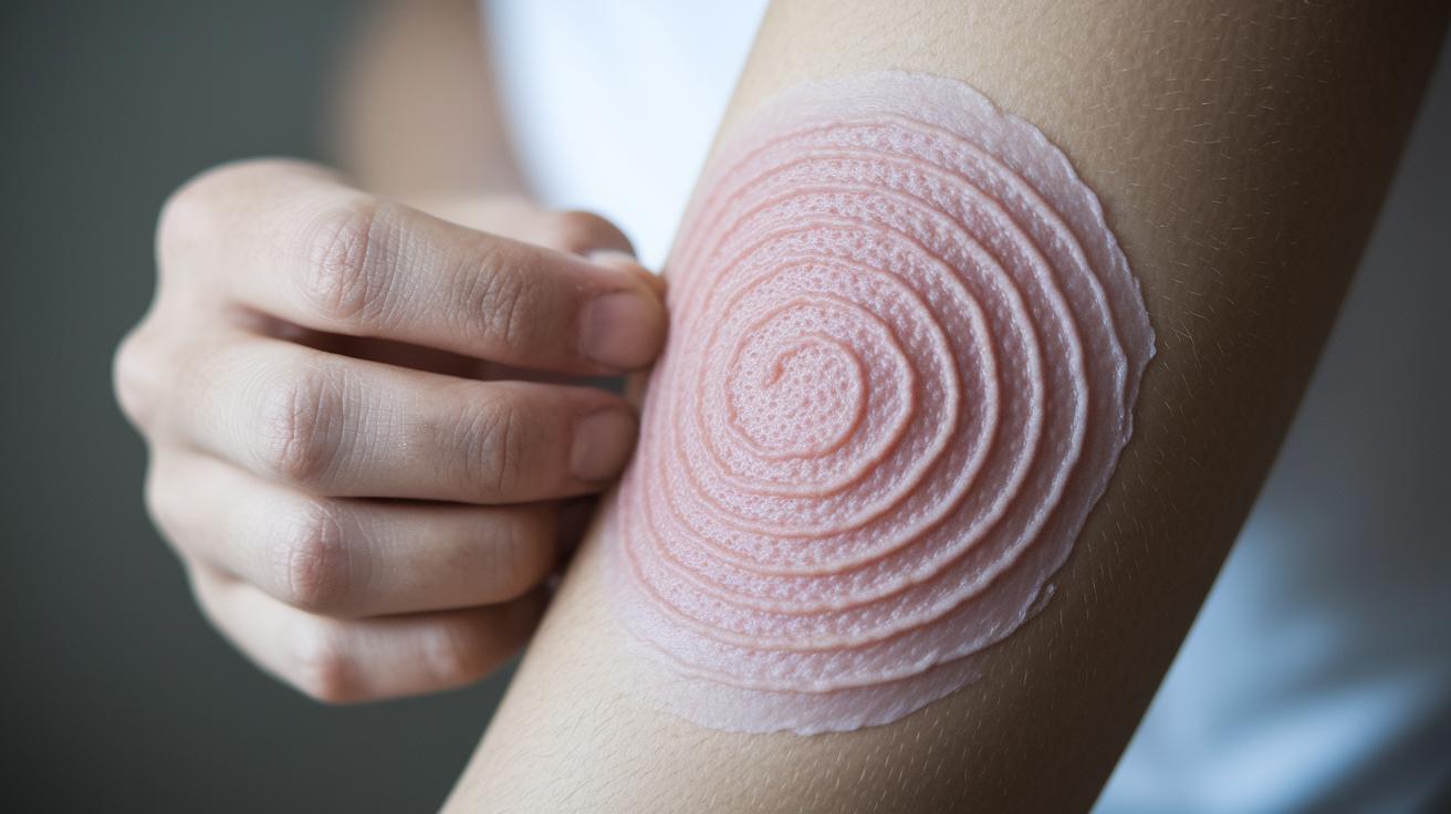
A crater-like skin lesion is a small bump with a round, sunken center. The center is filled with a hard, rough plug made of keratin (a protein in your skin, hair, and nails). It looks like a tiny volcano with a hardened cap.
Keratoacanthoma, or KA, is one such lesion that grows quickly. It usually measures between 1 and 2 cm. KA appears as a smooth bump with a central keratin plug that gives it a crater look. It tends to form on skin that gets a lot of sun, like your face, head, neck, arms, and legs. Sunlight, chemical exposure, a weak immune system, certain medications (like BRAF inhibitors), HPV infections, or minor skin trauma can all trigger KA.
Earlier, KA was called molluscum sebaceum. It mainly affects people aged 50 to 69 and happens twice as often in men, especially those with lighter skin (Fitzpatrick I-III). This change in naming shows how both environmental and personal factors shape its appearance and direct how it is treated today.
Imaging Techniques for Crater-Like Lesion Assessment
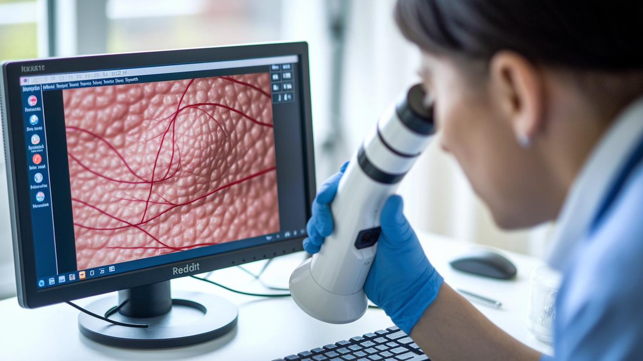
Noninvasive tests help doctors check skin marks that look like craters. Dermoscopy gives a close view of a lesion so you can see its keratin plug (a buildup of a skin protein), blood vessel patterns, and clear edges. This helps your doctor decide if the lesion might be a keratoacanthoma (a fast-growing bump) or needs more attention. In tricky spots, like under the nail, an X-ray can spot tiny bone changes (osteolysis or bone loss) that affect treatment and biopsy plans.
| Imaging Modality | Diagnostic Role |
|---|---|
| Dermoscopy | Shows keratin plug, blood vessel details, and lesion borders |
| X-Ray | Detects bone loss under nail lesions |
When these imaging methods are combined with a full clinical exam, your doctor gets a clearer picture. Small differences seen on dermoscopy can indicate whether you need quick treatment or just careful monitoring. This approach helps you get safe and effective care.
Causes & Risk Factors of Crater-Like Skin Lesions
Crater-like skin lesions happen when your skin is hurt by different triggers. Too much sun, harsh chemicals, and other factors can weaken your skin's ability to heal. When your skin's repair system is overloaded or your immune system is low (the body remains less able to fight off problems), these small depressions can appear.
Here are some common reasons you might see these lesions:
- UV radiation from sun exposure
- Direct contact with chemical irritants or cancer-causing substances
- Immune suppression from medications or diseases
- Use of specific treatments like BRAF inhibitors
- Genetic mutations (changes in p53 or H-Ras)
- Infection with human papillomavirus (HPV)
- Local trauma or having had surgery on the skin
Sometimes, these factors work together. For example, too much sun can speed up damage from genetic changes, increasing your risk of these skin depressions.
Differential Evaluation of Crater-Like Skin Lesions
Quick take: If you notice crater-like skin spots, check them now. Some changes need quick care.
Triage:
• Call emergency services now if you see rapid growth, bleeding, or severe pain.
• Get same-day care if the lesion changes fast, has uneven edges, or becomes swollen.
• Track the spot’s size, shape, and color daily and tell your clinician if it worsens.
When you spot a crater-like lesion, look closely at its shape, how fast it’s growing, and any changes in color or border. Your skin’s history also matters when deciding if it is harmless or needs more attention.
Some lesions may point to squamous cell carcinoma. This type can grow quickly and change texture, which is a sign it might be serious. Basal cell carcinoma usually shows as a shallow, ulcer-like spot that may not seem as aggressive but still should be watched. Amelanotic melanoma can be trickier because it may look like an irregular crater without dark spots, which is a red flag. On the other hand, molluscum contagiosum usually shows a dimple in the middle along with several similar bumps, suggesting it is a common viral change. Infections from fungus or bacteria can cause local erosion with red, inflamed skin.
A close-up tool called dermoscopy (a special skin exam) helps by showing detailed views of the lesion’s borders, tiny blood vessels, and surface scales. This method, along with your medical history, aids your doctor in making the right call quickly.
Diagnostic Approach & Biopsy for Crater-Like Skin Lesions
If you notice a crater-like spot on your skin, your doctor will start with a careful exam. They will look at the spot's shape, size, and texture. Using a tool called dermoscopy (a magnified skin exam), they can see details that help decide where to take a sample.
Your doctor will choose the biopsy method based on how deep the spot is and how it looks. Often, they will do an excisional biopsy, which means they remove the whole lesion with a 4 mm border around it. For smaller or deeper spots, a punch biopsy is better because it takes a small core of tissue without removing too much skin. If the lesion only affects the top layer of your skin, a shave biopsy is used to gently remove the surface. When the spot is near or under a nail, your doctor may first use imaging to check if the bone is involved before any tissue is taken.
It is very important to use the right margins and handle the sample with care. Removing a precise 4 mm border helps make sure there is enough tissue for a clear diagnosis and lowers the chance of missing any abnormal cells. This careful process helps your doctor plan the best treatment for you.
Treatment & Management of Crater-Like Skin Lesions

The standard treatment for crater-like skin lesions is surgical removal. Your doctor will usually cut out the lesion with a 4 mm margin to ensure that enough tissue is available for a proper lab analysis. When appearance and function are important, Mohs micrographic surgery is often used. This approach removes one thin layer at a time, saving as much normal skin as possible while confirming the diagnosis with lab tests.
If surgery is not the best first step, other options might work. For smaller lesions, a quick, office-based method like electrodessication (using electric current to burn the lesion off) with curettage (scraping the lesion) is sometimes used. In other cases, your doctor may try creams or injections directly into the lesion, although these are less common because there is not much evidence to support their use yet. For patients with many lesions or lesions in difficult spots, radiotherapy, which uses focused radiation to control the growth, can be a beneficial treatment.
The outlook after treatment is generally good, with a low risk of the lesion spreading. Rare complications, such as perineural spread (when the lesion moves along nerves), can happen. That is why regular skin checks are key. Monitor your skin closely and contact your doctor if you notice any new changes or if the lesion comes back.
Monitoring & Follow-Up for Crater-Like Skin Lesions
Quick take: Keep up with regular check-ups and home exams to catch any changes early.
If you see these red flags, contact your doctor immediately:
- Rapid recurrence of the lesion
- Persistent sores
- Unusual nerve symptoms (numbness or tingling)
At your doctor’s office, schedule skin exams every 6–12 months. These short visits let your doctor look closely at the area where the lesion was removed and check for any new crater-like depressions or signs of nerve issues.
At home, learn to check your skin regularly and protect it from the sun. Watch for any red flags such as the lesion coming back quickly, sores that do not heal, or new feelings like numbness. If you notice any of these, contact your doctor right away for a re-evaluation. This routine care helps you detect any problems early and work closely with your provider to keep your skin healthy.
Final Words
In the action, this guide broke down crater like skin lesion basics. It explained what these lesions look like, why they form, and how to tell them from other bumps. We highlighted imaging, diagnostic steps, and treatment options so you can quickly understand next steps.
Each section showed clear, step-by-step advice for safe self-monitoring and the need for prompt evaluation if red flags appear. Stay informed and proactive, and keep a positive focus on your skin health.
FAQ
What do crater-like lesions on skin look like?
Crater-like lesions typically appear as dome-shaped growths with a central, hardened plug. Pictures show round, sunken areas that you might see in conditions like keratoacanthoma.
What is keratoacanthoma and what do its pictures show?
Keratoacanthoma is a fast-growing skin lesion measuring about 1–2 cm. Its images reveal a dome-shaped nodule with a central keratin plug on sun-exposed skin areas.
How does keratoacanthoma differ from squamous cell carcinoma?
Keratoacanthoma grows rapidly with a distinct central plug, while squamous cell carcinoma appears more irregular and invasive. Both can look similar, so a biopsy is key for accurate diagnosis.
Is keratoacanthoma a type of cancer and is it dangerous?
Keratoacanthoma is generally considered non-cancerous despite its fast growth. It rarely spreads but still needs evaluation to rule out any malignant features.
What causes crater-like lesions on the skin?
Crater-like lesions can be triggered by UV radiation, chemical irritants, immune suppression, targeted medications, and local trauma, all of which can damage skin cells and lead to lesion formation.
Can skin cancer look like a crater?
Yes, some skin cancers may mimic crater-like lesions by forming depressed, irregular areas. Prompt evaluation is important to determine the exact nature of the lesion.