
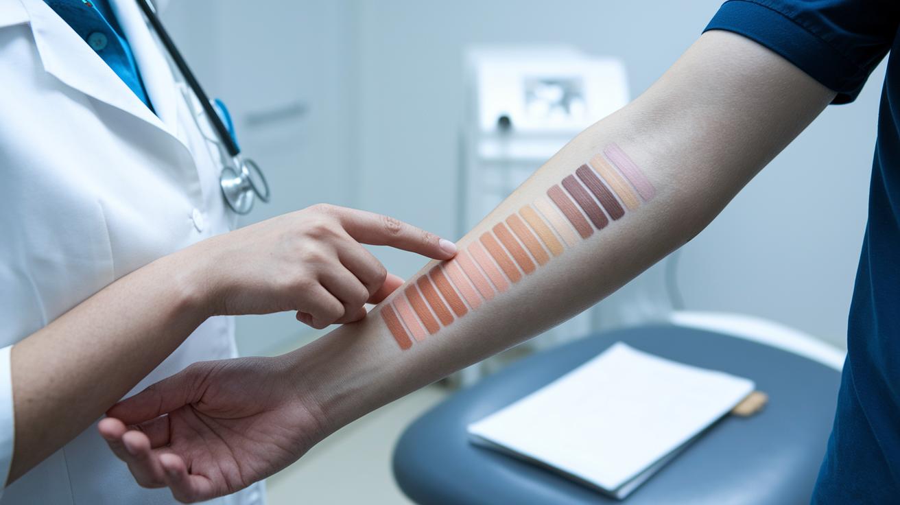

Quick take: Skin spots can be normal, but fast changes or discomfort may need a doctor’s look.
Have you seen a dark or light spot on your skin and wondered why? This is called dyschromia (color changes in the skin), which can show up as subtle shifts or clear marks. Most spots are harmless, but if they change quickly or cause discomfort, you should get checked. In this article, we explain common terms like hyperpigmentation (skin darkening) and hypopigmentation (skin lightening) in clear, simple language so you know when to seek care.
Skin Discoloration Medical Term: Precise Health Clarity
Quick take: Skin color changes can happen for many reasons. Most cases are not emergencies, but you should track any changes closely.
Triage Box:
• If you notice rapid color changes with pain, swelling, or signs of infection, seek same-day care.
• If the discoloration comes on suddenly or you feel unwell, contact your clinician immediately.
• Otherwise, monitor the area and note any changes over a few weeks.
Dyschromia is a general term for any unusual change in your skin's color. This word covers both darker and lighter spots and helps doctors group these issues without guessing an exact cause right away.
Hyperpigmentation happens when your skin makes extra melanin (the natural pigment that gives skin its color). This leads to dark patches or spots. In contrast, hypopigmentation is when your skin makes less melanin, causing lighter areas. For example, acne can leave behind brown marks (postinflammatory hyperpigmentation), while a condition like vitiligo creates pale patches.
Some conditions have clearer names. Melasma tends to cause even, brown patches in areas exposed to the sun and is linked to changes in hormones. Lentigo shows up as small, well-defined brown spots from long-term sun exposure. Vitiligo causes white patches because the immune system attacks the cells that produce melanin.
Classification of Skin Discoloration Conditions
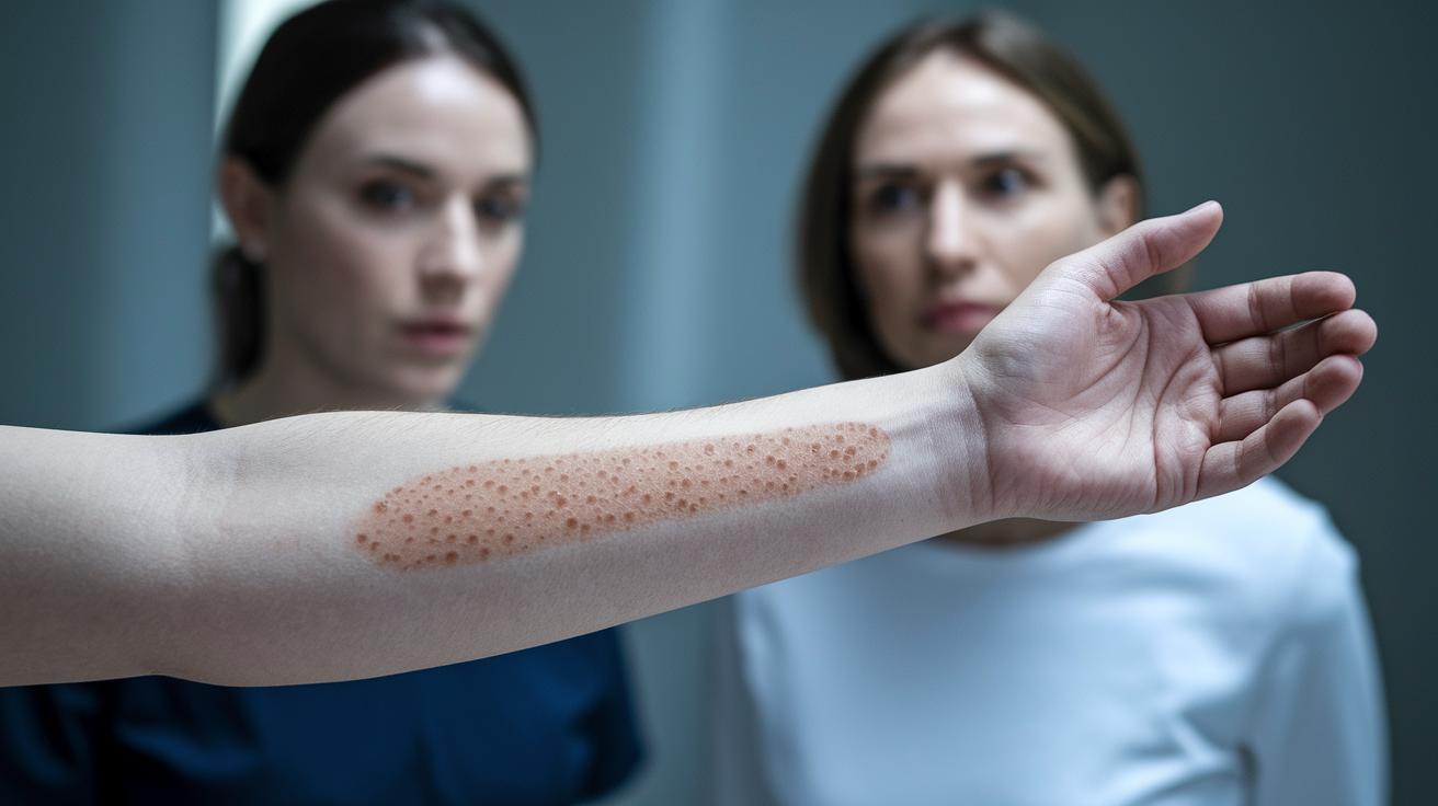
Quick take: Recognizing the type of skin discoloration helps your doctor choose the best care plan.
If you notice sudden changes, pain, fever, or a rapid spread in discoloration, call your doctor immediately.
Skin discoloration can occur for several reasons. Knowing which type you have can speed up diagnosis and guide treatment.
• Hyperpigmentation: Dark patches from extra melanin (the skin pigment).
• Hypopigmentation: Light spots from reduced melanin.
• Mixed dyschromia: Areas with both dark and light changes, leading to an uneven look.
• Vascular discoloration: Color changes (red or blue tones) due to blood vessel issues.
For example, dark patches on sun-exposed skin may suggest hyperpigmentation. Lighter spots might indicate conditions like vitiligo or changes after inflammation. A mix of dark and light areas calls for careful review, and red or blue hues could signal blood flow or inflammation concerns. Talk with your doctor to decide if further tests or treatment are needed.
Skin Discoloration: Hyperpigmentation Disorders, Terminology and Examples
Extra pigment happens when your skin makes more melanin (the natural color in your skin) than usual. This can show up as spots that are darker than your normal skin tone. Sun exposure, changes in hormones, or skin injuries can trigger this extra pigment. Doctors look at these differences to decide the best way to care for your skin.
Melasma
Melasma shows up as even brown patches on your face. It is linked to hormones and can get worse with sun exposure. You might notice melasma if you have been pregnant or use birth control. Its smooth, blended look sets it apart from other skin changes.
Solar Lentigo
Solar lentigo appears as small, neat brown spots on areas like your hands, face, and arms, where sun exposure is common. These spots are a sign of long-term sun damage. Under a microscope, doctors see extra pigment in the skin cells. This condition is harmless even though it shows sun damage.
Postinflammatory Hyperpigmentation
Postinflammatory hyperpigmentation comes as brown marks after your skin has been irritated by conditions like acne or eczema. These spots usually show up days or weeks after the injury and can stick around for months. They are more common if you have darker skin. Noticing when the spots appear after skin injuries can help in understanding and treating them.
Sunspots
Sunspots are flat, brown patches often called age spots. They develop slowly due to long-term sun exposure and can vary in size. Wearing sunscreen and protective clothing every day helps lower your chance of getting them. Your doctor may also recommend creams or light therapies to treat them.
| Condition | Medical Term | Key Features |
|---|---|---|
| Melasma | Melasma | Even brown patches tied to hormones and sun exposure |
| Solar Lentigo | Solar Lentigo | Small, neat spots from long-term sun exposure |
| Postinflammatory | Postinflammatory Hyperpigmentation | Brown marks that show up after skin injury or inflammation |
| Sunspots | Sunspots | Flat brown patches from chronic sun exposure |
Skin Discoloration: Hypopigmentation Disorders, Terminology and Examples
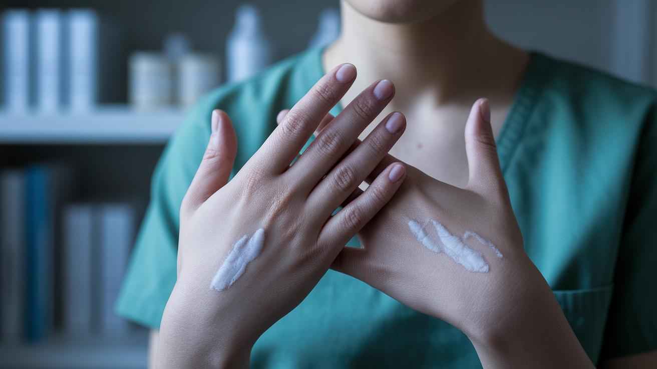
Melanin gives your skin its color. When your body makes less melanin, lighter spots may appear. This drop in pigment can happen when the skin is injured or when your immune system mistakenly harms the melanocytes (cells that produce melanin). These lighter patches are known as hypopigmentation and can be a sign that something else is going on.
Vitiligo is one common example. In vitiligo, your immune system attacks your melanocytes by mistake. This causes clearly defined, light patches that can appear anywhere on your body. Vitiligo affects about 0.5–2% of people, and the spots usually show up in similar areas on both sides of your body. They may grow in size over time, so it’s important to note if they change quickly.
Albinism is a genetic condition that happens because of a problem with the enzyme tyrosinase. This defect means you are born with less melanin, resulting in a pale skin tone along with light hair and eyes. Another example is postinflammatory hypopigmentation. This happens when an injury or inflammation, such as a burn, acne, or eczema, causes lighter skin in the affected area.
Underlying Causes and Pathophysiology of Skin Discoloration
Quick Take: Skin color changes can happen because of your genes, hormones, and what your skin is exposed to.
Triage Box:
• Call emergency services now if you suddenly develop severe pain, fever, or rapid swelling along with color changes.
• Seek same-day medical help if you notice new, painful, or spreading patches.
• Watch and self-manage if the changes are gradual and not linked with pain or other concerning signs.
Your genes decide a lot about how your skin makes its color. Sometimes, a change in an enzyme like tyrosinase (a helper protein for making melanin, the natural pigment) leads to less melanin and lighter spots. Hormones also play a big role. Shifts in estrogen and progesterone can make your melanocytes (cells that produce melanin) more active, which may cause melasma (brown patches on the face). These genetic and hormonal factors work side by side. For instance, if your family has a history of pigment changes, you might see color variations during puberty or pregnancy when hormones change.
Sunlight, skin injuries, and chemicals add another layer to the picture. UV rays (sunlight) boost melanin, often forming dark patches or age spots. Inflammation from injuries, acne, or eczema can either ramp up melanin production or damage melanocytes. This reaction sometimes leaves a dark mark after a cut, burn, or breakout, a change known as postinflammatory hyperpigmentation. Topical creams, such as corticosteroids or chemical bleaches, further affect skin color. Prolonged corticosteroid use may lighten the skin, while misused bleaching products might cause uneven patches. Together, internal factors and outside influences shape how your skin looks.
Clinical Assessment and Diagnostic Methods for Skin Discoloration
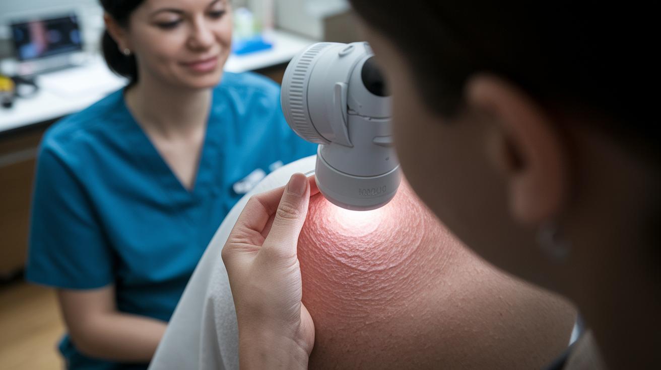
Quick take: Skin discoloration is usually checked with simple tools, but rapid or odd changes need prompt attention.
Triage Box:
- If you notice sudden changes in your skin’s color or texture, call your clinician now.
- If lesions look irregular or start changing quickly, seek same-day medical advice.
- If you have routine discoloration, schedule a regular check-up.
Dermoscopy is a noninvasive exam that uses a special magnifying tool to show details of your skin’s color patterns. This test helps your clinician see the pigment network (the natural design of skin color) and spot small differences between skin spots. A Wood’s lamp is another easy test. It shines ultraviolet light on your skin to show whether an area has less pigment (lighter skin) or no pigment at all (missing color). For example, if you have uneven skin tone, a dermoscopy can reveal subtle changes that you might not see on your own.
When a spot looks very different or changes quickly, a more detailed test is needed. In that case, a clinician may take a small skin sample (biopsy) to check the cells under a microscope. This process, called histology, counts pigment cells (melanocytes) and reviews how pigment (melanin) is spread through the tissue. This careful check helps confirm the diagnosis and rule out skin cancer.
For more about how doctors move from checking symptoms to making a diagnosis, visit how doctors go from symptoms to diagnosis.
Treatment and Management Protocols for Skin Discoloration
When your skin has extra pigment, clinicians often use creams and procedures together. You might be given topical treatments such as hydroquinone (2–6% strength for dark spots), retinoids (vitamin A creams), or corticosteroids (anti-inflammatory creams) to slow melanin (skin pigment) production. These creams work slowly to fade dark areas and can be combined for stronger effect.
Laser therapy is another option. Lasers like the Q-switched Nd:YAG or fractional lasers break up pigment clusters. In many cases, these lasers clear 50–80% of the pigmented spots. You may also consider cosmetic treatments such as chemical peels or microdermabrasion to remove the top layers of skin. Note that peels and microdermabrasion might bring back pigment if not used carefully.
Choosing the right treatment is very important. Your clinician will review your skin type and the severity of your discoloration to pick a safe and effective method. For example, a controlled laser can target deeper pigment while creams work best on surface areas.
When skin loses pigment, as happens with conditions like vitiligo, repigmentation methods are used. Narrowband UVB phototherapy (light treatment) can restore color in about 60% of cases. The treatment might cause mild redness or irritation in some people. Often, phototherapy is paired with topical medications to even out your skin tone when there are both dark and light spots.
To keep your skin looking even, regular check-ups with your clinician are key. Here are some tips to help manage and maintain improvements:
By keeping an eye on your skin and following these steps, you can fine-tune your care and work toward lasting improvement in skin discoloration.
When to Seek Professional Evaluation for Skin Discoloration
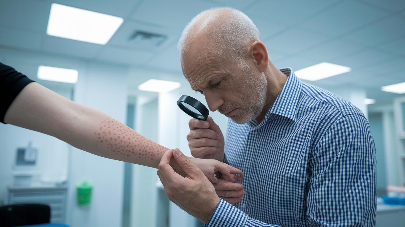
Quick take: Sudden or weird changes in your skin color can mean something is wrong, act quickly if you see these signs.
If you have any of these danger signs, call your doctor now:
- Spots with jagged or uneven edges.
- Lesions that change size, shape, or color fast.
- Areas that itch a lot, hurt, or bleed.
- Changes after surgery or injury, especially if you have a history of skin issues.
Sometimes a new or changing stain on your skin is more than just a spot. When you see these rapid changes, your skin could be under stress. That might signal a more serious problem.
Your doctor might check your skin using simple tools like the Fitzpatrick scale (a way to rate your skin type) or a score for melasma (a measure of skin discoloration). These tools help track how bad the change is and decide when you need another check-up.
Monitoring your skin regularly helps catch any worsening signs early, so you can get treatment fast and protect your health.
Final Words
In the action, we broke down key medical terms like dyschromia and skin discoloration medical term definitions to help you spot puzzling color changes. We outlined the classification into hyperpigmentation, hypopigmentation, mixed dyschromia, and vascular discoloration, then discussed examples like melasma and vitiligo. We also looked at causes, diagnosis, treatment options, and when to seek urgent care. Keep these points in mind as you monitor your skin changes. Stay proactive and positive, you have the tools to manage your symptoms safely.
FAQ
What is dyschromia and what does it include?
Dyschromia refers to any abnormal skin pigmentation. It includes conditions with increased melanin like hyperpigmentation (dark patches) and reduced melanin like hypopigmentation (light areas).
How do hyperpigmentation and hypopigmentation differ?
Hyperpigmentation occurs when extra melanin forms dark spots, whereas hypopigmentation results from a loss of melanin causing lighter patches on the skin.
What common skin discoloration conditions should I know about?
Common conditions include melasma (brown, hormone-driven patches), solar lentigo (sun-induced spots), vitiligo (autoimmune depigmentation), and postinflammatory changes following skin injury.
What are the underlying causes of skin discoloration?
Skin discoloration can be driven by genetic factors, hormonal imbalances, ultraviolet exposure, inflammation, or chemical influences that disrupt normal melanin production.
When should I seek professional evaluation for skin discoloration?
If you notice rapidly changing or irregularly bordered lesions, associated pain, bleeding, or persistent discoloration after injury, contact a healthcare professional immediately for evaluation.