

Quick take: Neurofibroma bumps are soft, skin-colored growths that are usually harmless.
If you notice any of these signs, call your doctor:
• The bump grows rapidly
• It becomes painful or tender
• It changes color or shape
Neurofibromas are skin bumps that most often cause no harm. They are soft and blend in with your skin, so spotting one can be confusing and even a bit scary. In this article, we explain what these bumps are, why they appear, and which changes you should watch for. This information can help you know when it’s time to check in with your healthcare provider.
neurofibroma skin lesion: Bright Medical Insights
Quick take: Neurofibroma skin lesions are soft bumps from nerve tissue that usually do not cause harm.
Triage Box:
- If you notice sudden severe pain, rapid growth, or a change in color, call emergency services now.
- If the bump begins to hurt or looks different quickly, see your doctor soon.
- For steady, small, and harmless bumps, you can monitor them at home.
Neurofibroma skin lesions are non-cancerous growths that develop from the covering of nerves. They feel like small, soft, skin-colored lumps under your skin. Many people compare them to a tiny button you might feel during a routine check.
These bumps often start during puberty, though some appear at birth. They might show up as one spot or as several small nodules that grow slowly over time. A child may have one tiny lesion that becomes easier to feel later, or an adult might notice a new bump that wasn’t there before.
Neurofibromas are linked to a genetic condition called NF1 (Neurofibromatosis type 1), which affects about 1 in 3,000 people. If one parent carries this gene, you have a 50% chance of inheriting it. This condition can affect anyone, though it is seen a bit more often in males. Knowing your family history is important.
These tumors are almost always harmless. Doctors only suggest removing them if they cause discomfort, rapid changes, or significant cosmetic concerns.
Classification of Neurofibroma Skin Lesions: Cutaneous, Subcutaneous, and Plexiform
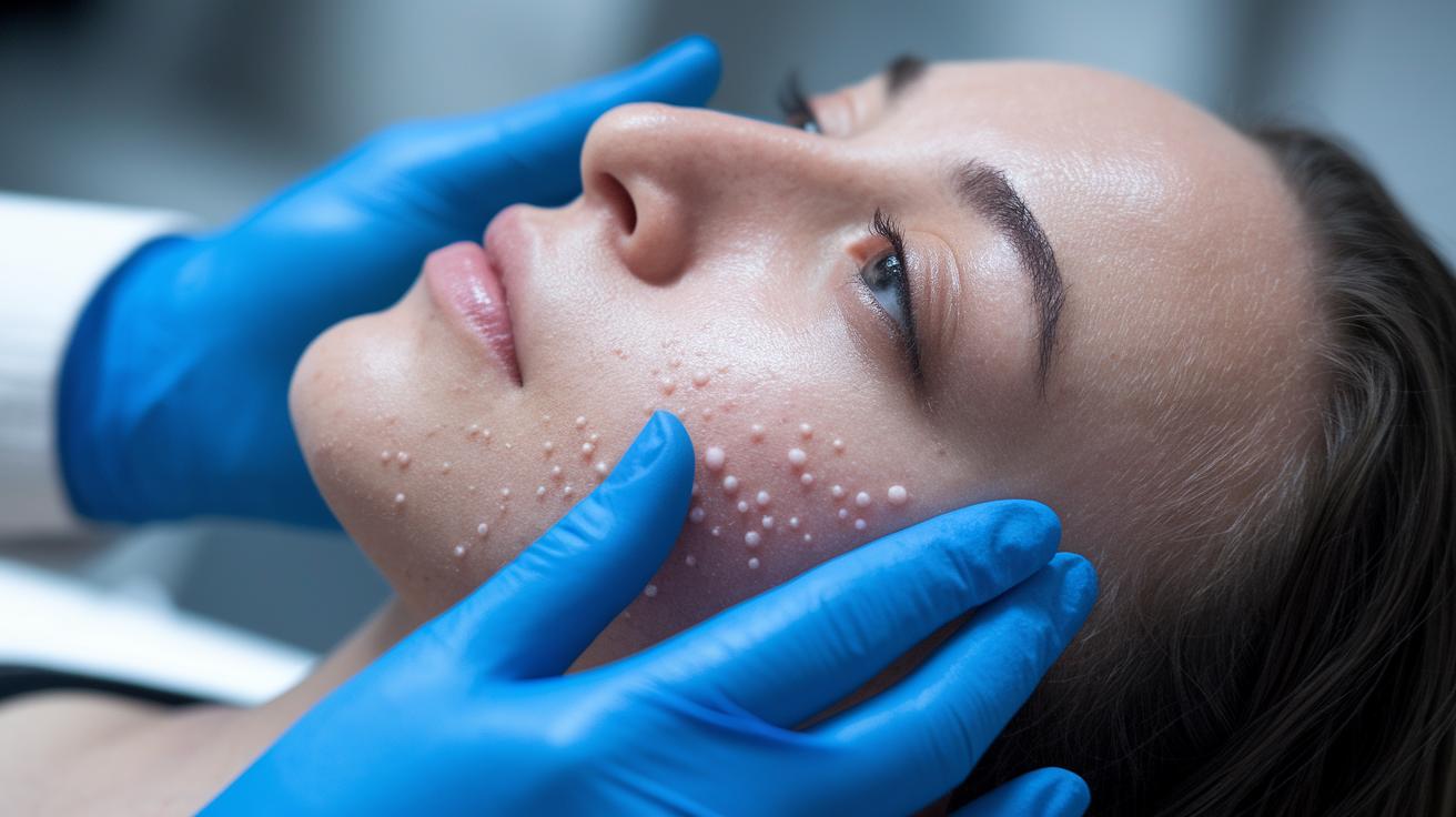
Neurofibromas are bumps that form on or under your skin. They may show up as a single lump or several little nodules. One bump often means a cutaneous type. Several bumps can be a sign of an inherited condition like NF1.
Cutaneous Neurofibromas
Cutaneous neurofibromas grow in the top layer of your skin (dermis). They feel soft, are skin-colored, and stick out like small buttons. You might notice a tiny, raised bump on your arm during a check.
Subcutaneous Neurofibromas
Subcutaneous neurofibromas sit deeper under your skin. They may feel firm and can cause mild tenderness or nerve discomfort when pressed. Though they are less visible, they might be more annoying because of their depth.
Plexiform Neurofibromas
Plexiform neurofibromas spread over multiple nerve bundles. They are usually present at birth and can expand over time. This type is more complex and may lead to noticeable swelling in the area.
| Type | Location | Key Features |
|---|---|---|
| Cutaneous | Upper skin (dermis) | Soft, skin-colored, small bumps |
| Subcutaneous | Under the skin | Firm, deeper, may hurt when pressed |
| Plexiform | Along nerve bundles | Spreads out, often present at birth, can enlarge |
Diagnostic Approach for Neurofibroma Skin Lesion
Quick take: Neurofibromas are soft, benign skin bumps that may signal NF1 when paired with key skin marks.
Triage Box:
If you notice these urgent signs, seek care immediately:
- The bump grows rapidly
- New pain or tenderness develops
- The color or shape of the bump changes
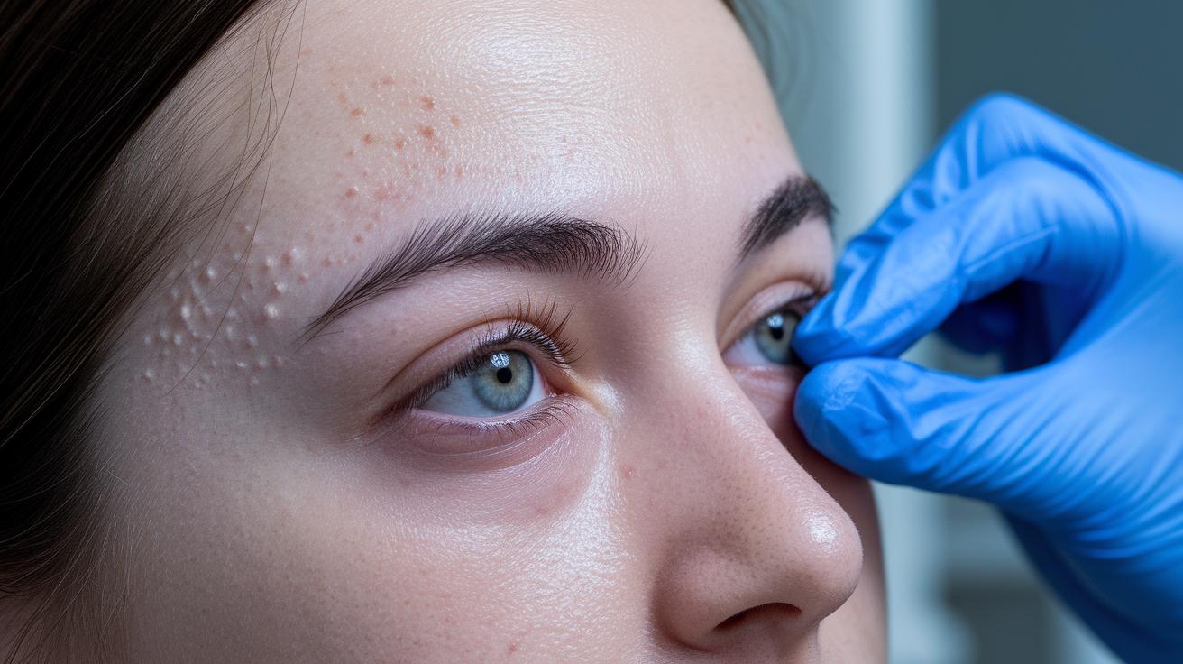
Clinicians use clear criteria to diagnose neurofibroma lesions when NF1 (a genetic condition) is suspected. They first look for café-au-lait spots (flat, light brown marks that appear by age 3) and freckling in the armpit or groin (typically seen between ages 3 and 5). A family history of NF1 further supports the diagnosis.
Physical Examination
During the exam, the doctor carefully inspects the skin and gently presses around the area to feel for soft, skin-colored nodules. They check the size, firmness, and how easily the bump moves under the skin. This helps determine if the bump feels like a typical neurofibroma.
Dermatological Criteria
In addition to examining the nodule, doctors look at the rest of the skin. They search for flat marks (macules) and small freckle-like spots in skin folds. These clues support the diagnosis and help set neurofibromas apart from other growths.
Biopsy Confirmation
A biopsy, in which a small tissue sample is taken for lab analysis, is reserved for bumps that look unusual or change quickly. The lab examines the cells under a microscope to confirm that the lesion is benign. This step rules out other types of tumors and confirms that the lesion is a neurofibroma.
Visual Characteristics of Neurofibroma Skin Lesion
Neurofibromas feel soft and are the same color as your skin. They are easy to press and may be round or a bit uneven, which makes them different from firmer bumps.
A small dimple can appear in the center when you press softly, and the edges gradually blend with the surrounding skin instead of standing out sharply. For example, a neurofibroma has a smooth feel, while a dermatofibroma feels firm and raised.
People with NF1 may also show other skin marks like café-au-lait spots (light brown patches) or freckles. These signs can help when you compare images in a skin lesion gallery.
Treatment Strategies for Neurofibroma Skin Lesion
Quick take: Most neurofibromas are harmless. You only need to worry if the bump becomes painful, grows quickly, or looks different.
Triage Box:
• If you experience severe pain, rapid growth, or disfigurement, seek urgent medical care.
• For any concerns about changes in the bump, contact your doctor right away.
Most neurofibromas are not dangerous. Your doctor may suggest regular check-ups to watch the bump unless it causes pain or grows faster than normal.
If the bump starts hurting, becomes disfiguring, or grows quickly, your doctor might recommend surgical removal. During this simple procedure, your doctor will numb the area with a local anesthetic and clean the skin thoroughly. Then, a small cut is made around the bump, which is carefully removed, and the cut is closed with fine stitches.
After surgery, you will need to keep the area clean and covered with a sterile bandage. Watch for signs of infection such as redness or swelling. Simple home care like this helps prevent any complications.
Your doctor will schedule follow-up appointments to check the wound and ensure the bump does not come back. This helps keep you safe and ensures a steady recovery.
Genetic and Inheritance Factors in Neurofibroma Skin Lesion
Quick take: NF1 is passed down from a parent and often shows up as noticeable skin spots and lumps early on, while NF2 usually causes tumors in the brain or spine without obvious skin changes.
Triage Box:
• If you spot new or changing skin spots or lumps, call your doctor.
• If the lesions grow, hurt, or change quickly, seek same-day care.
NF1 comes from a change in one gene and follows an autosomal dominant pattern. This means if one parent has the gene change, you have a 50% chance of inheriting it. In more than 85% of neurofibromatosis cases, the NF1 gene is altered. Family history is key because these changes can show up as café-au-lait spots (light brown patches) and neurofibromas (lumps on the nerves) at a young age.
In contrast, mutations in the NF2 gene occur less often and lead to different tumor types. NF2 usually causes tumors in the brain or spinal cord (called schwannomas) rather than clear skin changes. This is why NF1-related lesions are more common and visible, while NF2 changes may not create obvious skin signs.
Recognizing these differences helps you and your doctor decide on the best genetic testing and follow-up care for your specific risk.
Warning Signs and When to Seek Help for Neurofibroma Skin Lesion
Quick take: Watch your non-cancerous skin lesion closely and act fast if it changes suddenly.
If you have any of these signs, call your doctor right away:
- The lesion grows quickly in just a few weeks.
- You feel steady or worsening pain.
- You lose feeling or strength (numbness or weakness) near the lesion.
- The skin starts to break open or bleed without a clear reason.
- The lesion changes in a way that affects how it looks or works.
In a severe emergency with serious symptoms, call 911 or visit the nearest emergency room immediately.
Final Words
In the action, this article reviewed benign, soft nodules and their varying types. We covered how these lesions appear, what signs to look for, and when to get prompted care. You learned about different presentations, diagnostic checks, and clear red-flag cues for monitoring changes. Use the checklists and trackers provided to note any shifts in size, pain, or appearance. Stay informed and proactive when managing your neurofibroma skin lesion for a brighter, safer path ahead.
FAQ
Q: What do neurofibroma images and appearances show, including under-skin features?
A: The neurofibroma images show soft, smooth, flesh-colored nodules that can appear on or just beneath the skin. They are benign nerve tumors that vary in size and subtlety.
Q: What is a plexiform neurofibroma?
A: A plexiform neurofibroma is a complex, congenital nerve tumor that involves multiple nerve bundles. Although benign, its diffuse growth can lead to functional issues and requires careful monitoring.
Q: How is neurofibroma treated?
A: Neurofibroma treatment mostly involves watchful waiting since these tumors are benign. Removal is considered only if the lesion becomes painful, disfiguring, or grows rapidly, and it is done with careful surgical excision.
Q: Is a neurofibroma cancer?
A: Neurofibromas are not cancer. They are benign tumors composed of nerve sheath cells and rarely, if ever, transform into malignant growths.
Q: What is a solitary neurofibroma?
A: A solitary neurofibroma refers to a single benign nerve tumor that appears as an isolated nodule. It differs from multiple lesions seen in inherited conditions like NF1.
Q: How can you stop neurofibromas from growing?
A: There is no proven way to stop neurofibromas from growing. The focus is on regular monitoring, and treatment is aimed at addressing symptoms when they interfere with daily life.
Q: What are the symptoms of a neurofibroma?
A: Neurofibroma symptoms often include soft, skin-colored nodules that are typically painless but may cause local discomfort or nerve-related irritation if they press on nearby tissues.
Q: Should neurofibromas be removed?
A: Neurofibromas are usually left untreated unless they become painful, grow quickly, or are cosmetically concerning. Removal is recommended only when these issues affect quality of life.
Q: What causes neurofibromas?
A: Neurofibromas arise from benign growth of nerve sheath cells, often linked to genetic mutations such as those seen in NF1, which increase the likelihood of developing these tumors.
Q: How does neurofibromatosis affect pregnancy?
A: Neurofibromatosis can influence pregnancy by potentially increasing the size or number of neurofibromas. Pregnant women with this condition should work closely with their clinician to manage any changes during pregnancy.