
Quick take: Rough, scaly spots on your skin can be early signs of sun damage that need a check-up.
If you notice any of these signs, call your clinician:
• Bleeding or pain from the spot
• A spot that grows quickly or changes shape
• New spots that appear suddenly
Small, rough patches on your skin might be more than just an odd mark. These images show the subtle signs of long-term sun exposure. The condition, called actinic keratosis (rough, scaly spots from sun damage), may look like uneven, weathered skin. Even tiny spots can signal chronic damage.
This visual guide is here to help you catch early changes before they worsen. Spotting these signs now and getting a professional review can protect your skin later.
actinic keratosis pictures: Clear, striking details
These photos show signs of sun-damaged skin. They display rough, scaly patches that appear on areas like your face, scalp, ears, arms, and hands after long periods of sun exposure. If you notice these changes, get a professional check to make sure nothing serious is happening.
Images in this gallery capture small differences in skin texture. The patches come in shades from flesh tone to pink or brown. For example, one photo shows a rough patch on the forearm that looks like a dry, uneven landscape with a bit of crusting. Remember: one actinic keratosis spot can be a sign of decades of sun exposure. Even small marks deserve attention.
Key features to note include:
• Rough, scaly texture
• Colors from light pink to deep brown
• Lesions appearing on sun-exposed areas such as the face, scalp, ears, arms, and hands
• Variations in scale thickness and irregular edges on close-up views
These clinical images help you quickly spot early signs of chronic UV damage. If you see similar rough, persistent patches on your skin, it may be wise to have them checked by a clinician.
Close-Up Actinic Keratosis Photographs: Texture and Color Variations
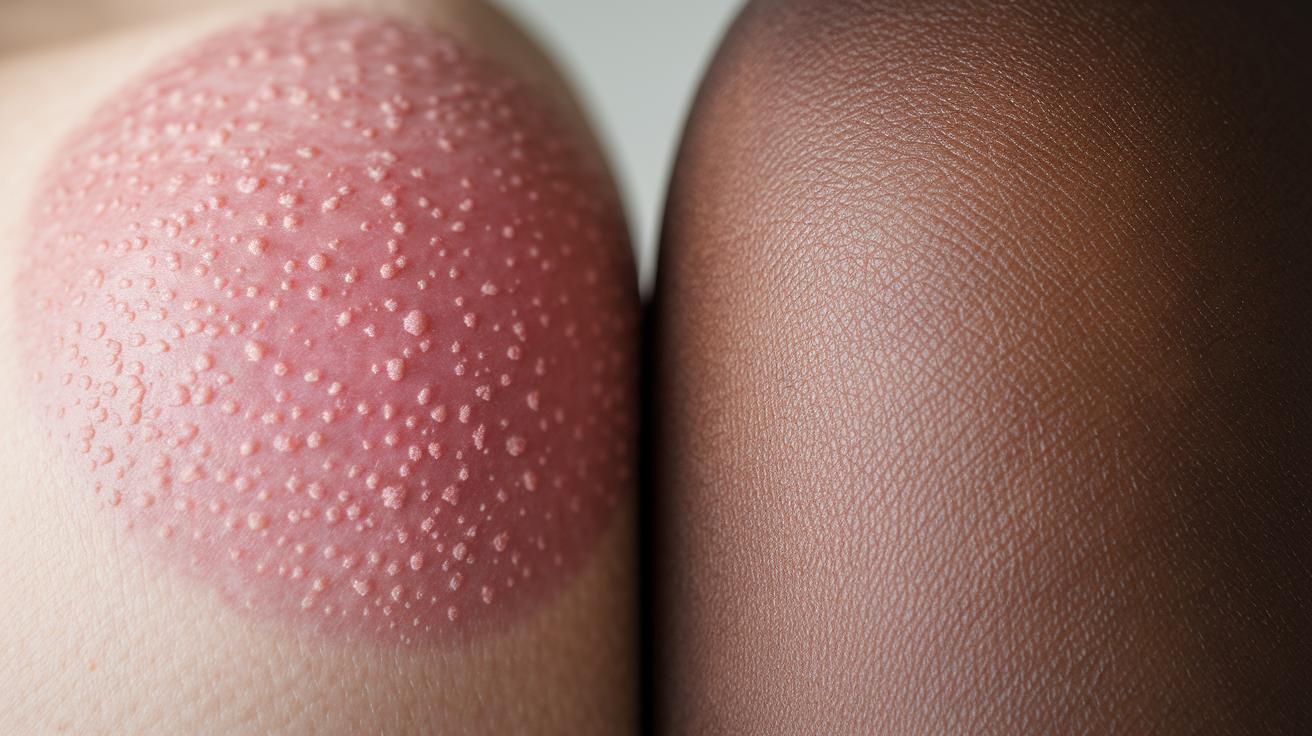
Close-up images let you see tiny details in skin affected by actinic keratosis (a pre-cancerous skin change). High-magnification photos show small bumps, flaky scales, and tiny dark spots that may hint at early cell changes. For example, you might notice a slight raised edge where normal skin meets an area with a color change.
These detailed photos reveal small texture differences:
- Noticeable roughness compared to the smooth feel of healthy skin.
- Color shifts that range from light pink to deeper brown.
- Fine scales and early keratin buildup that regular photos might miss.
Normal skin often looks even and uniform. In contrast, skin with actinic keratosis shows uneven borders and patchy scales. With high-magnification imaging, even early signs of skin change stand out clearly.
| Feature | Normal Skin | Actinic Keratosis |
|---|---|---|
| Texture | Smooth and even | Rough with tiny scales |
| Color | Uniform tone | Shifts from light pink to dark brown |
| Magnification view | Subtle details | Clear micro irregularities |
Sun-Damaged Patch Images vs Actinic Keratosis Photographs: Pictorial Comparison
Quick take: Uneven, flaky skin patches may signal actinic keratosis. If you notice rough patches with variable colors and borders, it’s time to check with your doctor.
If you have any of these warning signs, contact your dermatologist:
• Rough, uneven scales with crusting
• Color shifts from skin tone to pink or brown
• Irregular, not smooth, borders
Actinic keratosis shows a rough, scaly look with hints of crust that feel uneven. You often see different colors and jagged edges. In a side-by-side view, one image may show a rough, flaky lesion while the other shows a smooth, waxy patch. That difference tells you that actinic keratosis is not as uniform as some other skin spots.
Other skin conditions look different. For instance:
• Seborrheic keratosis is usually smooth and waxy.
• Eczema appears as red, inflamed, and itchy patches.
• Psoriasis has thick, silvery scales over reddened skin.
Remember, around 10% of actinic keratosis spots might turn into a more serious skin problem. That is why it’s important to look closely and talk with a clinician if you see these signs.
Imagine looking at two similar patches. One shows uneven and flaky textures, a red flag for actinic keratosis, while the other is smooth and likely harmless.
| Feature | Actinic Keratosis | Other Conditions |
|---|---|---|
| Texture | Rough, flaky scales with some crusting | Smooth and waxy (seborrheic keratosis); red and inflamed (eczema); thick and silvery (psoriasis) |
| Border | Irregular and uneven | Smooth and uniform |
Body Area Distribution: Actinic Keratosis Picture Guide by Location
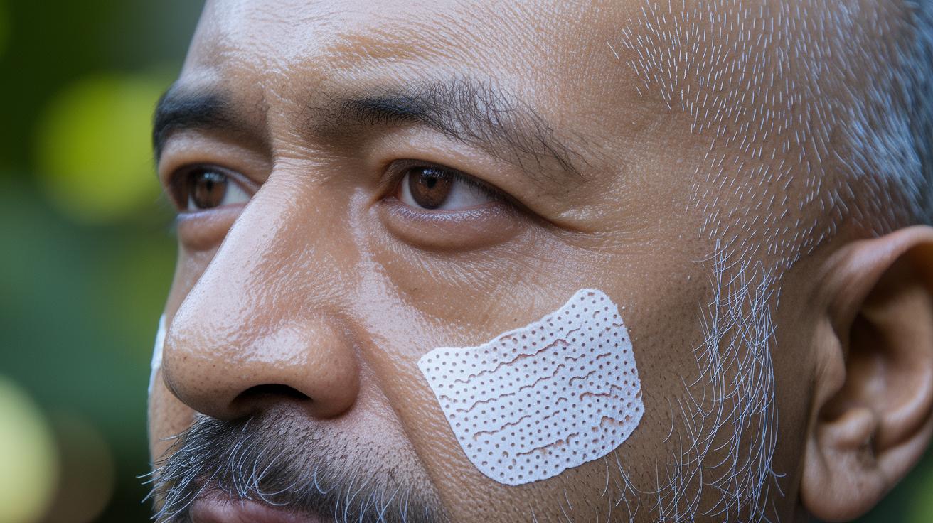
Actinic keratosis (sun-damaged skin patches) appears mostly on skin exposed to the sun. The images in this guide show where these spots usually form. For example, face photos often reveal patches on the cheek and the area between the eyebrows (glabella). Over 60% of these lesions occur there. The pictures help you recognize the rough, scaly texture that stands out against normal skin.
The scalp is another common site. In balding areas, about 20% of cases show dry, flaky patches along the hairline where the lesion meets healthy skin. Cases on the outer ear (helix) make up around 10%, highlighting irregular scaling. These visuals let you compare how actinic keratosis looks on less-covered areas that get a lot of sunlight.
Close-up images of the lips also provide key insight. While the lips are less often affected than the face, they can show color changes and very light scaling. Similarly, the backs of the hands and forearms display similar signs, with rough patches seen in about 10% of cases. These images clearly show how sun damage appears over time.
| Body Region | Estimated Frequency | Visual Characteristics |
|---|---|---|
| Face (cheek, glabella) | >60% | Rough, scaly patches with color variations |
| Scalp (balding areas) | ≈20% | Dry, flaky textured spots |
| Ears (helix) | ≈10% | Localized irregular scaling |
| Hands/Arms | ≈10% | Patchy lesions showing texture change |
Before and After Treatment Comparisons in Actinic Keratosis Pictures
These photos show how treatment can improve skin changes caused by actinic keratosis. Before treatment, you see rough, scaly patches with uneven edges. The spots can be flesh-colored, pink, or brown and often look raised and dry. These images highlight why starting treatment early is important.
Treatment options include creams like 5-fluorouracil and imiquimod, cryotherapy (freezing abnormal skin cells), and photodynamic therapy (using a light-sensitive agent plus light exposure). After treatment, the skin looks clearer. The patches become flatter, the scale fades, and the redness goes down. You usually see these changes in 2 to 8 weeks.
One set of images shows a lesion with heavy scaling before treatment. After cryotherapy, that same area is noticeably smoother and shows a softer, more even color. These before and after comparisons help both you and your clinician see how well the treatment worked.
| Before Treatment | After Treatment |
|---|---|
| Raised, scaly patches with uneven color | Smoother skin with reduced scale and redness |
Annotated Actinic Keratosis Pictures: Tracking Progression
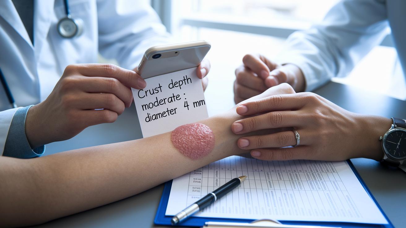
Quick take: These photos with notes help you track skin spots over time. If a lesion doesn’t start to heal after 2 weeks, call your doctor.
Doctors add simple notes to pictures of actinic keratosis (a skin spot that can turn into cancer) to show key details like size, texture, and crust (rough, scab-like area). Most lesions are between 2 and 6 mm wide. A note might read, "Crust depth: moderate; Diameter: 4 mm," meaning the lesion is a common size and has a rough patch.
These images are usually taken over 4 to 12 weeks. A time-lapse series shows if the spot is growing, staying the same, or shrinking. Changes in scale or color, or a lack of healing after 2 weeks, may signal that you need a biopsy (a small tissue test) to rule out a serious condition.
Key points to notice:
By reviewing these marked images, you can learn when a skin spot might need a professional check. Watching for changes helps you decide when to seek medical advice.
Self-Monitoring Visual Manual: Photographing and Tracking Actinic Keratosis Lesions
Quick take: Use clear, consistent photos to track your skin lesion. If a scaly patch does not improve in 2 weeks, call your doctor.
If you notice these signs, seek care immediately:
- The lesion grows quickly.
- The color or shape changes drastically.
- The lesion becomes painful or bleeds.
Here’s how to create a reliable photo record:
- Set up your space with steady, natural light. If natural light is low, use a lamp so your photo shows the lesion’s true color and texture.
- Place a ruler next to the lesion. This helps you and your doctor see any size changes over time.
- Take photos from different angles. Capture one shot at a slight angle to show the outline and another straight on to show depth and raised areas. For example, you might say, "I took two photos today – one at a slight angle and one straight on – to check the texture and size."
- Keep your photos consistent in angle and distance. This approach makes it easier to compare changes.
Make it a habit. Try taking a weekly photo diary. Write down the date and any changes like increased roughness, shifts in color, or size changes. If any scaly patch does not get better after 2 weeks, get a professional opinion right away.
Use this step-by-step guide to keep a clear record. For more details on tracking symptoms, you can review our full guide at https://thequickesttips.com?p=1347.
Final Words
In the action, this guide walked you through recognizing lesions with rough, scaly textures and distinct color shifts using vivid actinic keratosis pictures. The visual comparisons and marked differences between sun-damaged patches and treated lesions help you track changes over time. The clear step-by-step advice on self-monitoring aims to empower you to spot red-flag shifts early. Remember, reviewing these images may lead to timely professional advice. Stay positive and proactive with your skin health by keeping an eye on actinic keratosis pictures.
FAQ
What does actinic keratosis look like?
Actinic keratosis appears as rough, scaly, or crusty patches on sun-exposed skin, usually in colors ranging from flesh-toned to pink or brown. These lesions may resemble seborrheic keratosis, which are waxy and raised.
What is actinic keratosis in toddlers?
Actinic keratosis is uncommon in toddlers. It is mostly seen in older adults with prolonged sun exposure. If you notice unusual skin changes in a toddler, speak with a pediatric clinician.
What happens if actinic keratosis is left untreated?
Leaving actinic keratosis untreated can lead to persistent, sometimes worsening lesions. A small percentage may progress into squamous cell carcinoma (a type of skin cancer), so regular skin checks are advised.
What is the fastest way to get rid of actinic keratosis?
The fastest treatments include topical medications, cryotherapy, or photodynamic therapy. A clinician must evaluate lesion severity to choose the right treatment for your needs.
Where can I find pictures of actinic keratosis on different body areas?
Photos of actinic keratosis are available for various sun-exposed areas such as the face, arms, and legs, helping you compare texture, scale, and color variations with similar skin conditions.
How do before and after photos inform treatment progress for actinic keratosis?
Before and after photos help track treatment progress by showing changes in lesion texture, size, and color. Such images can demonstrate the reduction in scale and fading of redness after therapy.