
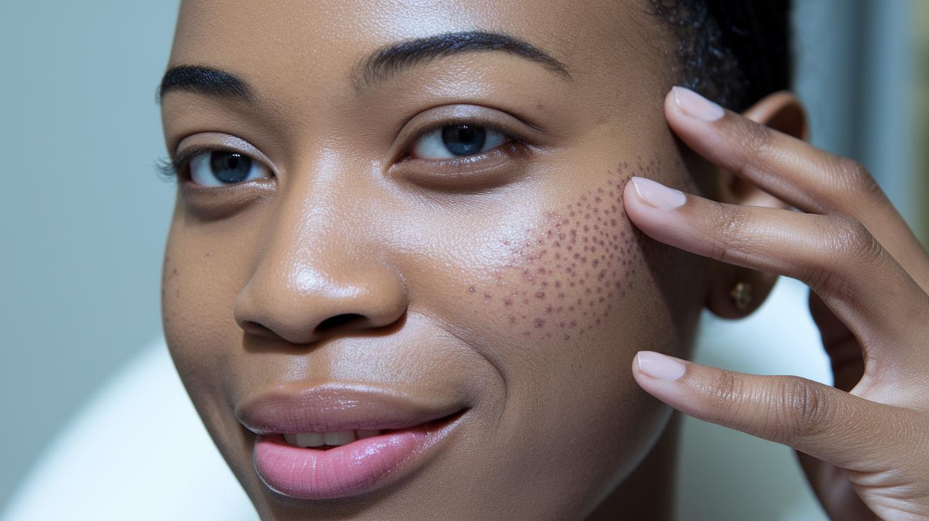

Quick Take: Even small changes in your moles can signal skin cancer. Watch them closely, and act fast if you see red flags.
If you notice any of these signs, call your doctor now:
• Moles with uneven or irregular borders
• Multiple colors or sudden changes in color
• Rapid growth or changes in size
• Bleeding or itchiness
Every day, sun exposure can make your skin change in tiny ways. Even small shifts in a mole may hide warning signals. With skin cancer cases on the rise, knowing what to look for helps you act fast and feel more confident about your health.
Keep an eye on your skin. Write down the date and any changes you spot with your moles. When in doubt, trust your gut and talk to a doctor for a checkup.
Your health matters, don’t wait to get help if you see these signs.
Recognizing Skin Cancer Lesions on the Face: Key Signs and Symptoms
Quick take: A new or changing mole on your face may be an early sign of skin cancer.
Triage Box:
• Red flags: If a mole starts bleeding, becomes very tender, or grows quickly, call emergency services now.
• Urgent concerns: If you notice a mole that has a mix of colors, uneven edges, or changes in size or shape, seek same-day care from your dermatologist.
• Monitor: If you see a spot that seems different but is stable, track its size, shape, and color changes and follow up with a clinician if needed.
Skin cancer strikes many Americans, about 1 in 5 by age 70. Your face gets lots of sun, which can lead to skin changes. Daily UV exposure (sunlight that reaches the skin) may trigger changes that could be early skin cancer.
Even tiny spots or subtle shifts on your face may signal trouble. Research shows that cases of melanoma (a serious type of skin cancer) might rise by 5.9% by 2025, and deaths could increase by 1.7%. That means any new or changing mole is a warning sign.
Keep an eye on these ABCDE features:
• Asymmetry: One side of the mole does not match the other.
• Border: The edges are irregular or jagged.
• Color: There are several or uneven shades.
• Diameter: The mole is larger than 6 mm (about the size of a pencil eraser).
• Evolving: The spot is changing in size, shape, or color.
Also watch for the Ugly Duckling sign. This means one mole or spot looks very different in color, size, or shape compared to others on your face. It’s normal to have many moles, but if one stands out, it deserves attention.
If you spot any of these signs, it’s wise to get an evaluation from a dermatologist quickly. Early detection and treatment can make all the difference.
Common Types of Facial Skin Cancer Lesions: Basal Cell, Squamous Cell, and Melanoma
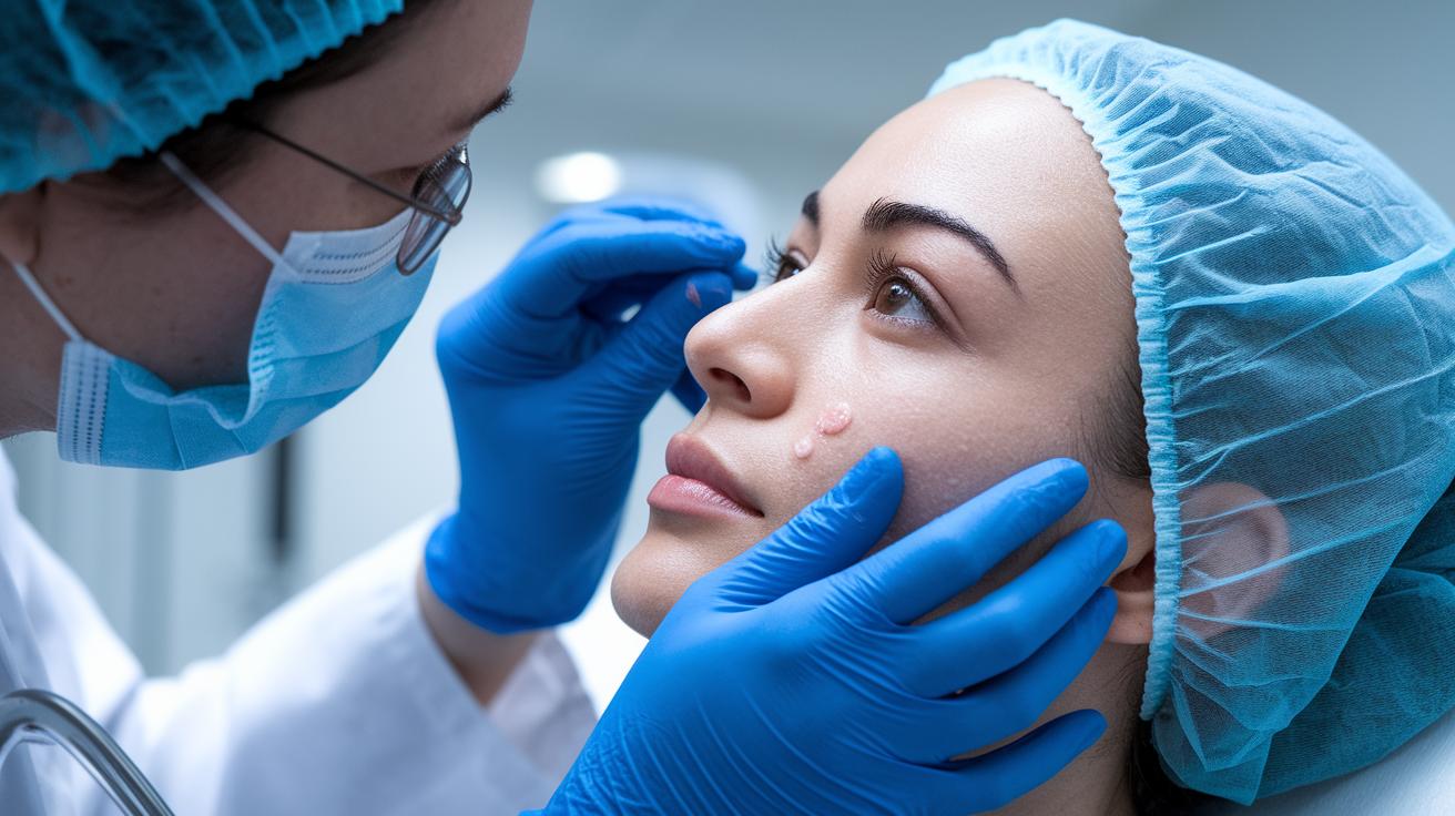
Quick take: If you see any new bumps, patches, or sores on your face that change or do not heal, get checked out right away.
Red Flags – Call emergency services if you notice:
- A sore that bleeds or doesn’t heal within a few weeks
- A bump that grows or changes shape and color
- Any lesion that looks different from the rest
Basal Cell Carcinoma
This type of skin cancer often appears as a firm, shiny bump or a rough, scaly patch on sun-exposed skin like your nose, cheeks, or forehead. You may notice it bleeding or forming a sore that just won’t close up. It usually grows slowly. Still, if you spot a bump or an unhealing sore, it’s important to have it evaluated by a healthcare provider.
Squamous Cell Carcinoma
Squamous cell carcinoma may show up as a small pink or red bump, a tender spot, or a scaly patch on areas like your ears, lips, or scalp. This lesion can feel sore to the touch. People with certain risks, such as those who have had an organ transplant, are more likely to get this type. If you find a spot that feels tender and does not go away over several weeks, please seek medical advice.
Melanoma
Melanoma is the most aggressive form of skin cancer and can be tricky to spot. It might look dark brown, black, light brown, or even pink. The lesion can be small or large and may have uneven borders. Changes in color or shape are key warnings. For example, if a mole on your forehead develops irregular edges or a mix of colors, it is a sign that you need to get it checked immediately.
Diagnostic Methods for Facial Skin Cancer Lesions
Quick take: Checking your face early for skin changes can lead to simple and effective treatment.
If you notice any rapidly changing, bleeding, or painful spots on your face, seek medical care immediately.
• Self-monitoring: Do monthly face checks using a mirror or body map.
• Urgent care: If a spot grows quickly or causes pain, see a dermatologist right away.
• Follow-up: Track any changes by taking pictures and noting details.
Regular self-exams help you spot small changes that might otherwise go unnoticed. When you see a new or evolving spot, a clinical exam is needed. Dermatologists use a tool called a dermoscope (which magnifies skin details) to look closely at the area. They also use high-resolution images, like digital dermatoscopic photos, to compare changes over time.
If a spot looks abnormal, your doctor may perform a biopsy. There are different methods to take a small piece of skin for testing under a microscope (this test is called histopathology). These methods include punch, shave, or excisional biopsy. Finding skin cancer early like this can result in cure rates over 90% when surgery is used.
Follow these five key steps:
- Check your face every month – Use a mirror or map to spot new or changing marks.
- Get a professional exam – A dermatologist will use dermoscopy to examine any worrisome spots.
- Document areas with photos – Taking clear pictures helps track changes over time.
- Have a biopsy if needed – Your doctor will choose the right method to sample the skin.
- Use tissue results to guide treatment – A detailed exam of the sample will help plan your care.
Risk Factors and Prevention for Skin Cancer Lesions on the Face
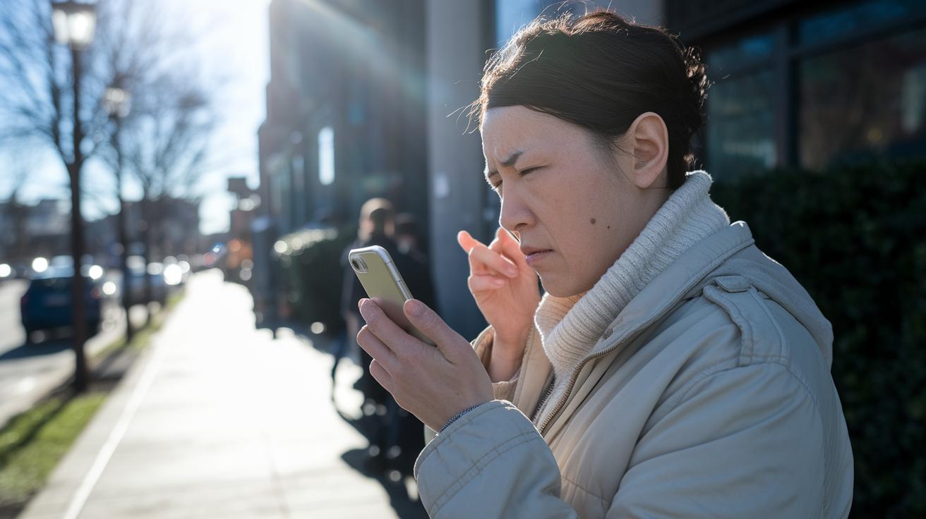
Quick take: Sun damage is the main cause of skin changes on your face, and early prevention can make a big difference.
If you notice any new, changing, or unusual spots on your face, call your doctor immediately.
Your face gets a lot of sun exposure. UV light from the sun and tanning beds can hurt your skin. People with fair skin, those who have had many sunburns, people over 50, or those with a family history of skin cancer are at higher risk. Even on cloudy days, UV rays can harm your skin, just like a bright sunny afternoon might catch you off guard.
To lower your risk, start by protecting your skin every day. Wear a wide-brimmed hat, sunglasses that block UV rays, and cover up with long sleeves when you can. Use a sunscreen labeled SPF 30 or higher, and reapply every 2 hours or after sweating. Try to stay in the shade during the busiest sun hours, and make time for regular skin checks with your clinician. These steps help prevent damage and catch any changes early, so you can act right away if something isn’t normal.
Stay alert and take care of your skin every day.
Treatment Options for Facial Skin Cancer Lesions
Quick Take: Surgery is the main treatment for facial skin cancers, though less invasive options work for early or surface lesions.
Triage:
• Red flags: If your lesion grows quickly, bleeds, or shows signs of infection (redness, pain, discharge), call your doctor immediately.
• Urgent: If your lesion starts affecting how your face works or looks, seek same-day medical advice.
• Monitor: If there are changes without red flags, keep track and follow up with your clinician.
Surgery is the standard approach for many facial skin cancers. For basal cell carcinoma (a slow-growing skin cancer) and squamous cell carcinoma (a common skin cancer), doctors often use surgical removal or Mohs micrographic surgery (a technique that removes the cancer layer by layer) to get every bit of the lesion while keeping as much normal skin as possible. Mohs surgery is ideal on the face because it is very precise. Removing the lesion completely can lower the chance it will come back and help ease concerns about looks or function.
For early, superficial lesions, treatments like cryotherapy (using very cold temperatures to destroy unwanted cells) or topical immunotherapy with imiquimod cream (which triggers your skin’s defenses) offer good alternatives. These methods focus on the abnormal cells and spare most of your healthy skin, making them a fitting option if you prefer a less invasive treatment or if the lesion was found early.
When surgery isn’t the best choice, other treatments come into play. Radiotherapy may be used when the cancer is aggressive or in a tricky spot on the face. New targeted therapies and anti-PD-1 immunotherapy trials, which boost your body’s natural ability to fight cancer, offer hope for more advanced melanoma. These systemic treatments are options when traditional methods don’t work or aren’t possible.
Post-Treatment Care and Cosmetic Reconstruction After Facial Lesion Removal

Quick Take: Follow your wound care steps and monitor for signs of infection. If you notice any red flags, call your healthcare provider right away.
Triage Box:
- If you have high fever (above 101°F), increased redness, warmth, swelling, or pus, call emergency services now.
- If you experience uncontrolled pain or sudden changes in your wound, seek same-day medical care.
- Otherwise, follow these self-care steps as advised.
After your facial lesion is removed, taking care of your wound is key. Clean the area every day with a gentle cleanser and apply a thin layer of antibiotic ointment as your clinician instructed. Cover the spot with a sterile dressing to help keep out germs and aid healing.
Once the wound has healed enough, you can take steps to reduce scarring. Use silicone gel sheets and gently massage the area as directed by your clinician. Also, avoid direct sun exposure on the healing skin to prevent dark or raised scars.
If your doctor recommends cosmetic reconstruction, there are several safe options. For small defects, a local flap closure uses nearby skin to naturally fill in the area. For larger wounds, a skin graft may be used to cover the space. Another option is contour restoration, which helps match the natural curves of your face. Your clinician will choose the best method based on the size and location of the lesion to protect both your look and facial function.
Finally, if you feel distressed about changes in your appearance, support groups or counseling can be very helpful. These resources offer practical advice and comfort as you continue your recovery.
Self-Examination and Ongoing Monitoring of Facial Skin Cancer Lesions
Quick take: Check your face every month and act quickly if you notice any changes.
Triage Box:
• If a mole or spot suddenly grows, changes shape, or shifts in color, call your healthcare provider immediately.
Every month, stand in good light with a mirror to study your face carefully. Look at your moles and any marks for small changes in size, shape, or color. If a spot seems to grow or look uneven, take note.
Use high-resolution photos to document any changes and mark them on a body mapping template (a simple chart that shows where each spot is located). Keep these records organized in a journal or an app so you can compare them side by side over time.
If you are at higher risk for skin cancer, schedule follow-ups with your dermatologist every 6 to 12 months. And if you notice any unusual change in a lesion, contact your healthcare provider right away.
Final Words
In the action of spotting warning signs, the article has shown you clear steps for identifying abnormal facial skin spots, understanding diagnostic methods, and exploring treatment options.
Following prevention measures and routine self-exams helps you catch skin cancer lesions on face early. Taking quick, practical steps and maintaining regular follow-ups empowers you to manage your health safely.
Stay focused and proactive. Your actions today can lead to a healthier tomorrow.