
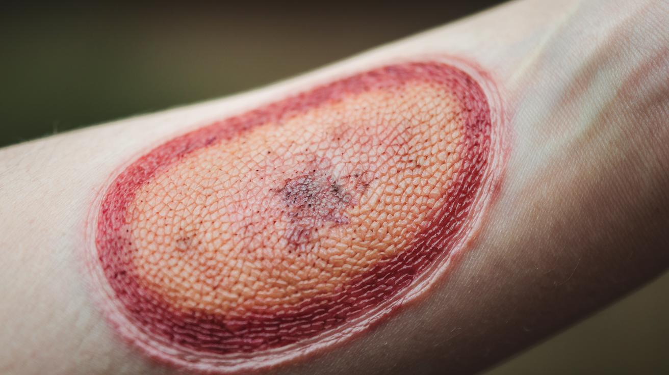

Quick take: A small skin spot can be harmless but sometimes may signal a problem that needs attention.
If you have any of these emergency signs, call emergency services now:
• The spot bleeds or oozes persistently.
• It grows rapidly or changes shape.
• It becomes very painful or itchy.
• You notice significant color changes.
Skin lesions, or spots on the skin, come in many shapes and colors. Some are primary lesions, which are the first visible changes, while others are secondary lesions that develop later or due to another issue. Knowing what to look for can help you act quickly if things change.
Here’s what to do:
• Check the spot regularly. Write down its size, color, and any changes.
• Note if you experience pain, itching, or bleeding from the area.
• If the lesion changes or you feel worried, reach out to your clinician.
This guide gives you clear details on skin lesion types and tips on when to seek care. Stay observant and protect your health by tracking any shifts in your skin spots.
Clinical Overview of Skin Lesion Descriptions
Quick take: Skin lesions can be harmless or a sign of something that needs quick attention.
Triage:
- Red flags: If a spot grows fast, changes shape or color, or starts bleeding, call your doctor immediately.
- Urgent care: If the lesion feels very painful or itchy, seek same-day care.
- Monitor: If nothing seems urgent, keep an eye on the lesion and note any changes.
A skin lesion means the skin looks or feels different than usual. It might look like a simple mole or freckle. It can also be a red rash or a blister from a virus.
Lesions come in two main types. Primary lesions start on normal skin. These include flat spots and raised bumps. Secondary lesions appear when a primary spot changes and leaves behind scales or crusts.
Many things can cause a lesion. Infections (germs causing illness), aging, injuries, allergies, long-term illnesses, and even family traits may be involved. The size, shape, color, and texture of a lesion depend on what is causing it and your skin type.
It can be worrying to see changes. If a lesion grows quickly, changes its shape, or alters its color, don’t wait. Call your doctor right away.
Doctors need clear details about what a lesion looks like. They note the size, color, and whether it feels hard or soft. For example, if a mole becomes uneven in just a few weeks, that is a sign to get checked.
Primary and Secondary Lesion Classification in Skin Lesion Descriptions
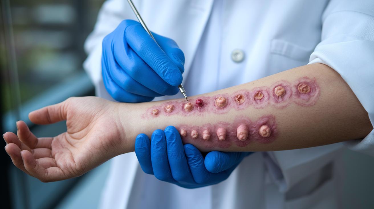
Primary lesions appear directly on healthy skin. They show up as spots or bumps that form without any earlier marks. You might see a flat discolored spot (macule), a small raised bump (papule), a deeper and larger bump (nodule), a tiny blister (vesicle), or a broad raised patch (plaque). For example, you might notice a red papule after a small irritation or a tiny flat freckle that has always been there. These features give important clues about the original skin issue.
Secondary lesions develop later when primary ones change. They often come about as the skin heals or after external interference. Common types include flaky patches (scales), hardened dried serum (crusts), areas where part of the skin is lost (erosions), deeper breaks in the skin (ulcers), and permanent marks (scars). If a papule turns into a crust, it might signal an infection or irritation that needs further checking. Recognizing these changes helps narrow down possible causes and guides safe care.
Visual Characteristics of Skin Lesion Morphology
Quick take: Noting skin lesion features closely can help you decide if you need to see a doctor.
If you notice any of these changes, call your doctor right away:
- The lesion grows quickly or gets larger.
- It shifts from one color to several hues.
- Its outline becomes jagged or uneven.
Common causes for these changes can be harmless or may need evaluation. Here’s how to check your lesion:
• Shape: Look at the lesion. It may be round, oval, or irregular. A round shape is often seen in a benign mole, while an irregular outline might call for closer observation.
• Size: Measure the lesion in centimeters. Many lesions are less than 1 cm, but some can be over 2 cm. A change in size over time should be noted.
• Color: Check the color. Lesions can appear pink, red, brown, black, or even lighter than the surrounding skin. A lesion with a uniform color is often harmless; mixed colors or sudden shifts can be a warning sign.
• Texture: Feel the surface. It might be smooth, rough, scaly, or crusted. A smooth surface might be less concerning than a rough, scaly one that may indicate a persistent irritation.
• Border and Surface: Note the edges and the surface level. Well-defined borders are generally less worrying than jagged ones. Also, see if the lesion is flat, raised, or indented, and gently feel if it is firm or soft.
Keep a record of these details and share them with your healthcare provider. This helps them understand whether the lesion is stable or if it needs further evaluation.
Lesion Configuration Types and Distribution Patterns
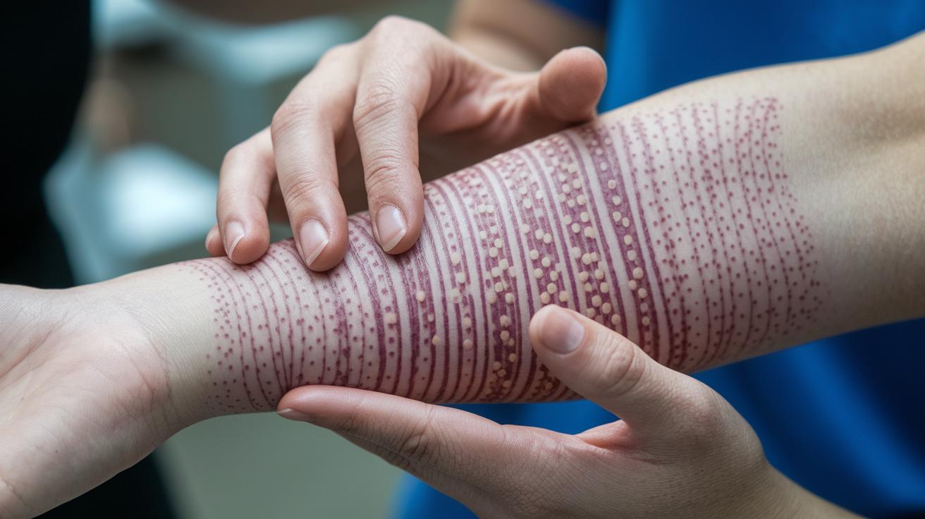
Quick take: The shape and spread of skin lesions can give important hints about what might be causing them.
Triage: If your skin changes suddenly, spread fast, or come with pain or fever, call your doctor now.
Skin lesions can appear in many distinct patterns. Some form rings with a clear center, which you may see with tinea corporis (a common fungal infection). Other times, lesions group closely together as seen in herpes simplex (a viral infection). When spots line up in a streak, it might be contact dermatitis, which happens when your skin reacts to an irritant.
Lesions that follow a nerve route, known as a dermatomal pattern, often point to shingles. Sometimes the spots form a netlike (reticular) pattern, as seen in livedo reticularis. Lastly, when small spots merge into larger patches or plaques, conditions like psoriasis or other long-term skin issues might be the cause.
It is important to note where these lesions appear. Are they in one spot, or do they cover larger areas? Do they show up evenly on both sides of your body or more on one side? Your doctor will use these details to decide if you need further checks or immediate treatment.
| Configuration Type | Description | Example Condition |
|---|---|---|
| Annular | Ring shaped with clear center | Tinea corporis |
| Grouped | Lesions clustered together | Herpes simplex |
| Linear | Arranged in a line or streak | Contact dermatitis |
| Dermatomal | Follows a nerve path | Shingles (herpes zoster) |
| Coalesced | Lesions merge into larger plaques | Psoriasis |
| Reticular | Netlike pattern | Livedo reticularis |
Distinguishing Benign vs Malignant Indicators in Skin Lesion Descriptions
Quick take: A stable, even spot is usually harmless, but any sudden changes call for a check by your doctor.
When your skin spot has a smooth shape, even color, a regular edge, and grows slowly, it is often a benign (harmless) lesion. Think of a small, uniform mole that stays the same over time, it is usually nothing to worry about.
However, some spots may show warning signs that need urgent evaluation. Watch for these red flags:
- Asymmetry: One half of the spot does not match the other.
- Border irregularity: Edges are uneven, jagged, or notched.
- Color variation: Different colors appear in one spot.
- Diameter greater than 6 mm: The spot is larger than 6 mm across.
- Evolution: Noticeable changes in size, shape, or color over time.
Also, a spot that does not heal or has an ongoing sore is a red flag.
If you see any of these changes, act now. Write down the details like size, color differences, or any other changes you notice. Share this information with your doctor at your next visit or sooner if the spot seems to be changing rapidly. Early attention can make a big difference in treatment and keeping your skin healthy.
Detailed Analysis of Specific Lesion Types in Skin Lesion Descriptions
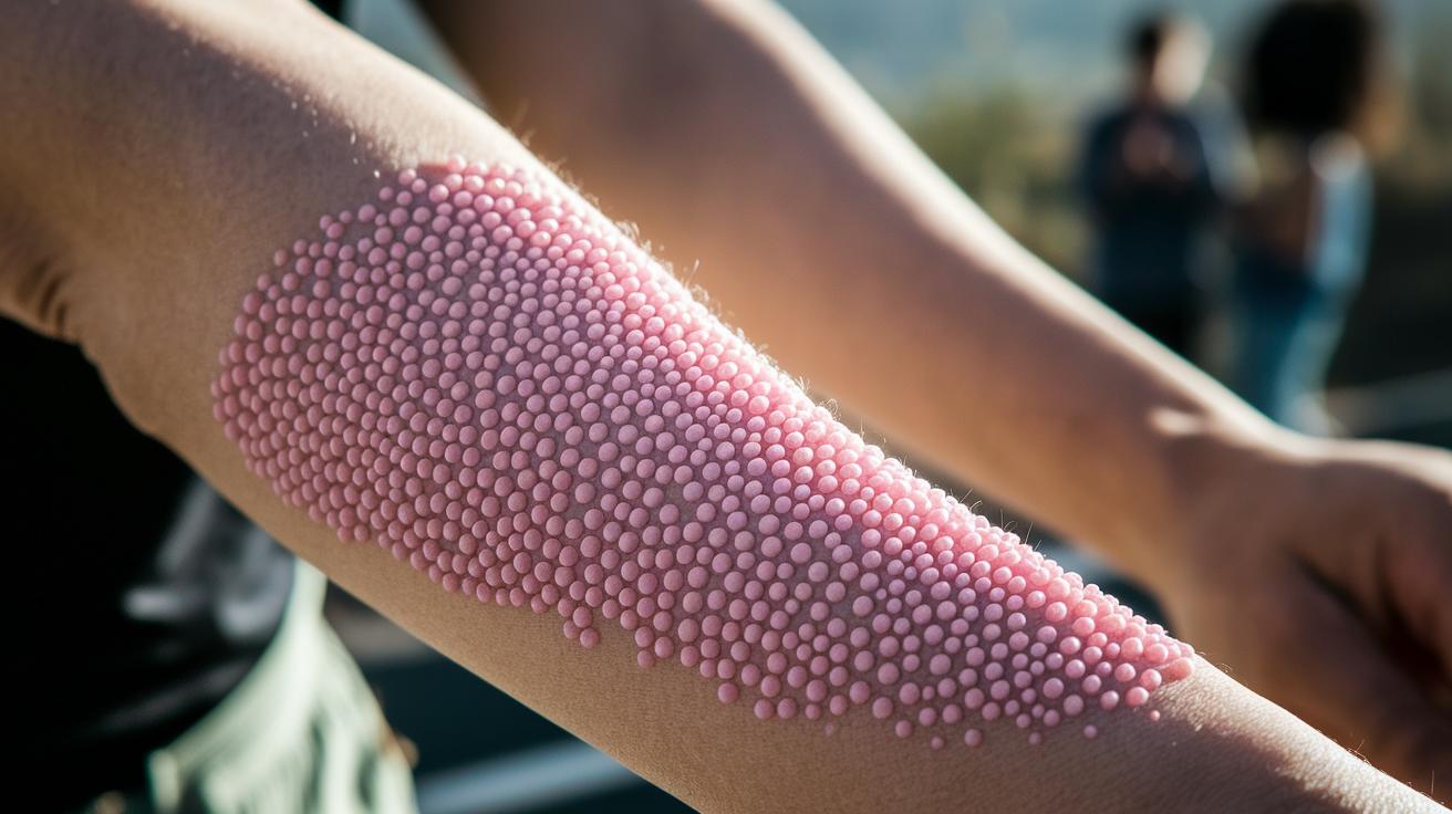
Actinic Keratosis Visual Markers
Actinic keratosis usually appears as small, rough, pink bumps on skin that has seen a lot of sun. They often feel a bit gritty when you run your finger over them. These bumps are common on the face, scalp, or arms because of long-term sun exposure. For example, you might notice tiny, rough patches on your forearm after a long sunny day that feel like fine sandpaper.
Seborrheic Keratosis Recognition
Seborrheic keratosis shows up as a waxy, stuck-on spot. You may see brown to black patches with small keratin cysts (tiny lumps of protein). They have a smooth surface that makes them look like they were pasted onto your skin. Imagine a spot that appears like an old piece of dried glue on your skin. Many people are surprised when they notice these marks and find that they stay the same over time.
Psoriasis Lesion Depiction
Psoriasis is marked by well-outlined red patches with a silvery scale (a thin layer that looks like white powder). These lesions appear on common spots such as the elbows or knees. The red patch has clear edges that stand out from normal skin. For instance, you might see a red spot on your forearm with a light, silvery covering that is very distinct.
Fungal Lesion Identification
Fungal skin infections often appear as round patches with a scaly border and a clear center. They usually come with itching that can be bothersome. The ring-like shape is a key clue for this type of infection. You might notice a circular red patch with raised, scaly edges and a clear middle. These signs help quickly point to a fungal skin issue.
Best Practices for Documenting and Reporting Skin Lesion Descriptions
Use a standard template when you record skin lesion details. A good template lists the exact spot, size, shape, color, edge, overall look, and any symptoms you notice. Keeping things the same way every time helps you track changes and share clear notes with other providers. For example, take a photo with a measuring tool to show the lesion's size and use a dermatoscope (a tool for a close-up look) to see surface details.
Stick to a common set of terms by using a dermatology glossary. This helps everyone use the same language and reduces mix-ups. Add these points to your SOAP notes (Subjective, Objective, Assessment, and Plan) for clear skin exams.
Here is a simple checklist to help you record lesion details consistently:
- Note the exact location of the lesion.
- Measure it and record its size.
- Describe the shape and color in plain language.
- Detail the edge or border.
- Watch for any changes in its overall look.
- List any symptoms the patient feels along with the lesion.
Using consistent notes helps you evaluate the skin carefully and spot changes that might need quick action.
Final Words
In the action, you’ve seen how a skin lesion description guides you through classifying and evaluating lesions. The post breaks down primary vs secondary lesions, visual markers like shape, size, and color, and lists red flags for potential malignancy. It also covers detailed analysis of common lesions such as actinic keratosis and psoriasis. This guide offers clear steps for documenting these features to help you decide on urgent care, monitoring, or simply tracking changes. Stay proactive and informed, knowing each detail brings you closer to safe, effective care.
FAQ
What do skin lesions pictures show?
Skin lesions pictures show images of skin changes from moles to rashes. They help you see differences in size, shape, color, and texture to compare normal and abnormal appearances.
What does a types of skin lesions chart include?
A types of skin lesions chart lists different lesion forms such as macules, papules, nodules, vesicles, and plaques. It serves as a quick guide for recognizing and categorizing skin changes.
What is a plaque skin lesion?
A plaque skin lesion appears as a raised, flat-topped area on the skin. It often covers a larger area than a papule and is commonly seen in conditions like psoriasis.
What is a papule skin lesion?
A papule skin lesion is a small, raised bump on the skin, usually less than 1 cm in diameter. It represents an initial change in skin structure.
What is a nodule skin lesion?
A nodule skin lesion is a firm, raised bump that is deeper and larger than a papule. It often measures over 1 cm and may require further evaluation.
What do benign skin lesions pictures depict?
Benign skin lesions pictures depict non-dangerous skin findings. They often show uniform color, symmetry, and regular borders, helping to differentiate harmless moles or freckles from worrisome changes.
What is a vesicle skin lesion?
A vesicle skin lesion is a small, fluid-filled blister that is usually less than 1 cm in diameter. It can be a sign of viral infections or allergic reactions.
What are primary and secondary skin lesions?
Primary and secondary skin lesions distinguish initial changes from those that evolve. Primary lesions arise directly on unaltered skin, while secondary lesions develop from changes like healing or infections.
How do you describe a lesion on skin and in pathology?
Describing a lesion involves noting its shape, size, color, texture, border, and elevation. In pathology, the description also includes microscopic features to assess underlying structural changes.
What are the three types of lesions?
The three types of lesions can refer to primary, secondary, and incidental findings. Primary lesions occur on normal skin, secondary lesions develop from primary changes, and incidental lesions appear during assessment.
What are the 10 primary skin lesions?
The 10 primary skin lesions include macule, patch, papule, plaque, nodule, vesicle, bulla, pustule, cyst, and wheal. These terms outline the basic presentations observed in skin examinations.